
|
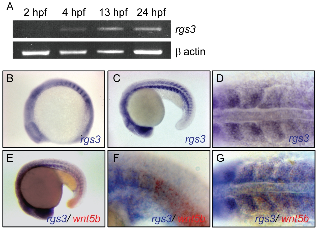
Fig. 1 Temporal and spatial expression of rgs3 throughout zebrafish development.
RT-PCR was used to determine the temporal expression of rgs3 from 0 hpf to 24 hpf (A). Whole Mount In Situ Hybridization was utilized to determine the spatial expression of rgs3 in 12 hpf (B) and 20hpf (C,D) wild type embryos. rgs3 and wnt5b double label in situ in18hpf embryos (E–G). Lateral (B,C,E,F) and dorsal (D,G) views illustrate that rgs3 is expressed in the developing somites (B–D) and posterior tail (C). At 18 hpf rgs3 expression is enriched in the posterior (caudal) portion of the developing somites (D). Co-localization of wnt5b and rgs3 was determined by double label WMISH with wnt5b (red) and rgs3 (blue) showing overlapping expression domains in the developing tail and somites (E–G). Sense probes (negative control) gave no specific hybridization signal.