
|
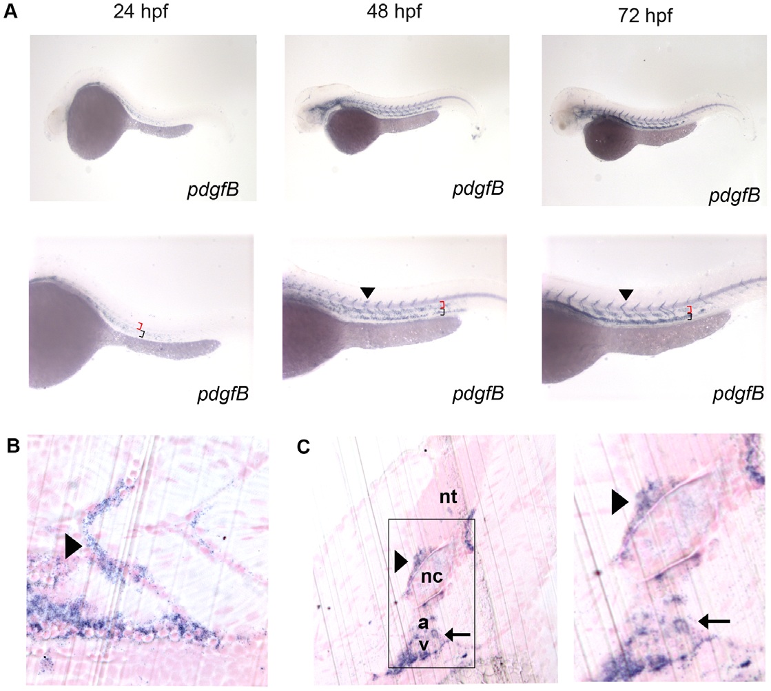
Fig. 3 Characterization of pdgf-b in the developing zebrafish embryo.
In situ hybridization of embryos at 24, 48, and 72 hpf (A) using an anti-sense probe against pdgf-b. pdgf-b was expressed at all time points in the head and tail vasculature (arrowheads indicate pdgf-b staining in ISVs, red brackets indicate the dorsal aorta, and black brackets indicate the posterior cardinal vein in A). Sagittal (B) and transverse (C) JB-4 sections of 72 hpf embryos analyzed with whole mount in situ hybridization using a probe against pdgf-b. pdgf-b expression was localized near the dorsal aorta, posterior cardinal vein and ISVs (arrow in C indicates pdgf-b staining in dorsal aorta and posterior cardinal vein while the arrowhead indicates staining in the ISV). Neural tube (nt), notochord (nc), dorsal aorta (a), posterior cardinal vein (v).