
|
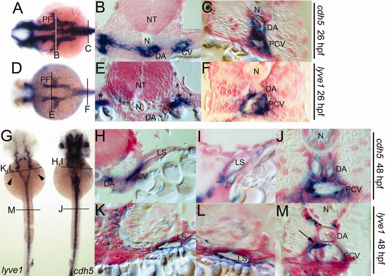
Fig. 4 A-M: Presumptive lymph sacs (LS) in transverse sections through pectoral fin bud region and trunk in 26 hours postfertilization (hpf) and 48 hpf embryos. cdh5 expression in 26 hpf (A-C), 48 hpf (G-J). lyve1 expression in 26 hpf (D-F), 48 hpf (G,K-M). LS expressing lyve1 in 26 hpf embryos (asterisks) were adjacent to the cardinal vein (CV) in the pectoral region, while in the trunk, lyve1 still labeled the posterior cardinal vein (PCV). Dorsal views at 48 hpf showed distinct lyve1 and cdh5 expression patterns within the whole embryo. Expression of lyve1 within the PCV appeared diminished and had become prominent within the inter-axial vessel space (arrow). PF, pectoral fin buds; N, notochord; NT, neural tube; arrowheads in G mark the positions of the pectoral fins.