
|
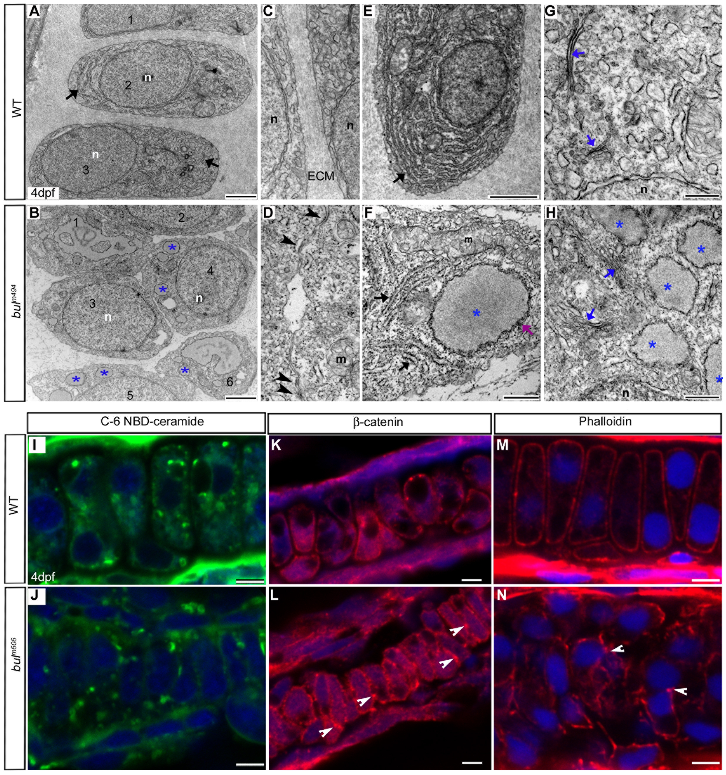
Fig. 3 Sec24D-deficient cartilage contains sparse ECM and chondrocytes with distended ER.
(A–H) Electron micrographs show mature wild-type chondrocytes (cells 1–3 in A) are separated by ECM and contain dense ER membranes (A, E, arrows). In bulm494 mutants (B,F,H), cartilage matrix is sparse and large amounts of electron-dense material accumulate in the rough ER (asterisks, cells marked 1–6 in B). Wild-type chondrocytes (C) are devoid of cell-cell contacts, whereas the majority of bulm494 mutant cells (D) contain adherens junctions (arrowheads). bulm494 chondrocytes (F) contain both normal ER cisternae (arrows) and swollen, ribosome studded, ER (asterisks). sec24d-deficient cells (H) contain smaller and disorganized Golgi complexes (blue arrows) compared to wild-types (G). (I,J) Single-pass confocal images of C6-NBD ceramide stained chondrocytes of wild-type (I) and sec24d/bulm606 mutant (J) embryos at 4 dpf. (K,L) Adherens junctions are marked by β-catenin (red) in bulldog Meckel′s cartilage (arrowheads), but are mostly absent in wild types (K). (M,N) Phalloidin (red) shows regular cortical distribution of polymerized actin in wild types (M), but uneven cellular distribution in bulm606 cartilage (arrowheads) (N). Nuclei are stained with TOPRO-3 (blue). Abbreviations: m, mitochondria; n, nucleus; ECM, extracellular matrix. Scale bars: 2 μM (A,B,E); 500 nm (F–H); 5 μM (I–N).