
Image
|
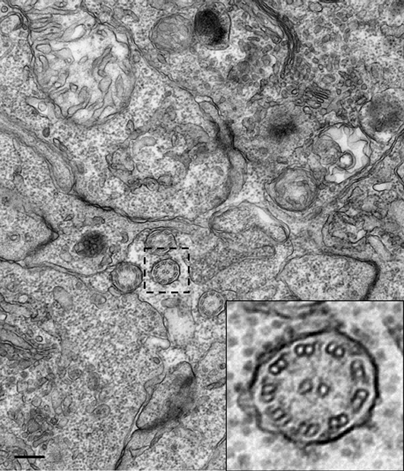
Figure Caption
Fig. S5 Cilia structure in the neural tube. Transmission electron microscopy image of cilia in the central canal of a 24 hpf embryo, showing a 9 + 2 structure. Boxed area indicates region magnified in inset. Scale bar: 25 nm.
Acknowledgments
This image is the copyrighted work of the attributed author or publisher, and
ZFIN has permission only to display this image to its users.
Additional permissions should be obtained from the applicable author or publisher of the image.
Reprinted from Developmental Biology, 341(2), Hong, E., Jayachandran, P., and Brewster, R., The polarity protein Pard3 is required for centrosome positioning during neurulation, 335-345, Copyright (2010) with permission from Elsevier. Full text @ Dev. Biol.