
Image
|
Figure Caption
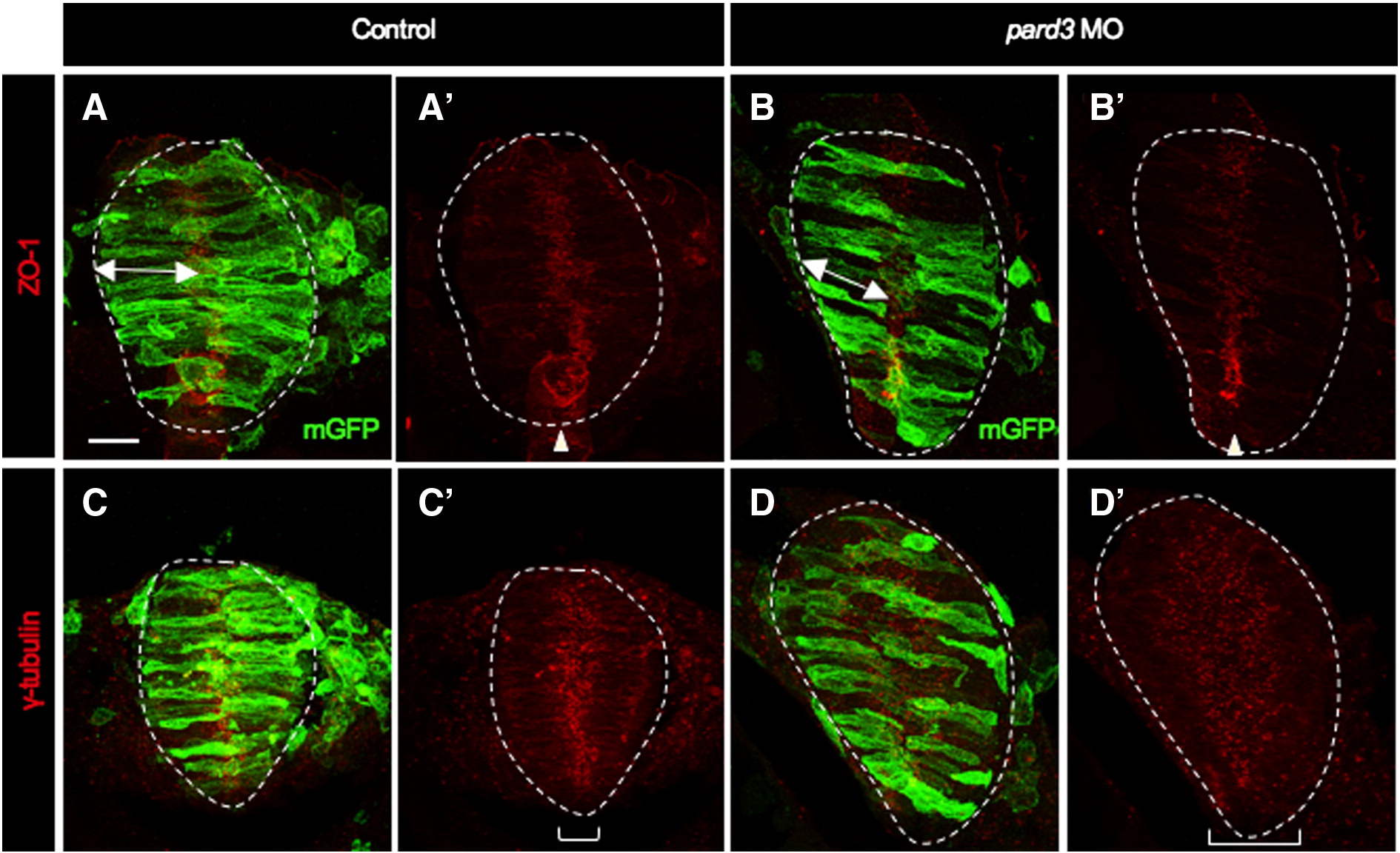
Fig. S4 Apico-basal polarity appears normal in pard3 MO-injected embryos. Cross sections through the hindbrain region of mGFP-injected control (A,A′,C,C′) and mGFP, pard3 MO co-injected (B,B′,D,D′) embryos at 12–13 som. Sections from the same embryos were immunolabeled using either anti-ZO-1 (A,A′,B,B′) or anti-γ-tubulin (C,C′,D,D′). White dotted line delineates the neural rod. Double arrowheads indicate length of cell. Brackets show the distribution of γ-tubulin throughout the neuroepithelium. Scale bar: 20 μm.
Acknowledgments
This image is the copyrighted work of the attributed author or publisher, and
ZFIN has permission only to display this image to its users.
Additional permissions should be obtained from the applicable author or publisher of the image.
Reprinted from Developmental Biology, 341(2), Hong, E., Jayachandran, P., and Brewster, R., The polarity protein Pard3 is required for centrosome positioning during neurulation, 335-345, Copyright (2010) with permission from Elsevier. Full text @ Dev. Biol.