
|
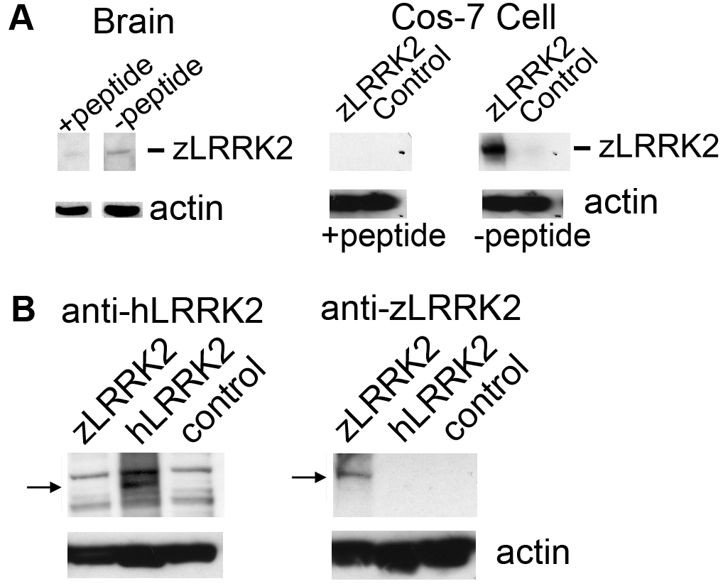
Fig. S10 Analysis of zLRRK2 antibody specificity. (A) Specificity of zLRRK2 antibody specificity was verified by western blot analysis. Positive signals could be largely blocked by pre-incubating the anti-zLRRK2 antibody with the neutralizing peptide (+ peptide, left panel). Similarly, the zLRRK2 signal was completely blocked by pre-incubating the anti-zLRRK2 antibody with the neutralizing peptide in the zLRRK2-overexpressing Cos-7 cells (+ peptide, right panel). (B) Western blot analysis showing the specificity of the zLRRK2 antibody against zLRRK2 in Cos-7 cell line. Human LRRK2 and zLRRK2 recombinant proteins were overexpressed in Cos-7 cells separately and detected by anti-hLRRK2 (left panel) and anti-zLRRK2 (right panel), respectively. A positive band (280 kD, arrow) was only detected in hLRRK2-overexpressing cells (hLRRK2, left panel), using anti-hLRRK2 antibody (NOVUS NB 300–268). No obvious band was detected in zLRRK2-overexpressing and untransfected control cells (zLRRK2 and control, left panel); On the other hand, a sharp band (arrow) was detected in zLRRK2-overexpressing cells using the anti-zLRRK2 antibody (zLRRK2, right panel), but not in hLRRK2-overexpressing and untransfected control cells (hLRRK2 and control, right panel). This indicates the anti-zLRRK2 antibody is specifically against zLRRK2 proteins.