
|
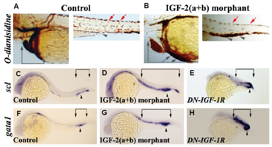
Fig. 4 Blood circulation is disrupted when IGF signalling is reduced. (A) Control morpholino injected embryos stained with O-dianisidine at 72 hpf show normal blood circulation. (B) IGF-2(a+b) morphant embryo with reduced circulating blood. Grey arrows indicate blood in the heart region, red arrows blood circulation in the intersomitic vesicles and grey arrowheads the intermediate cell mass. (C,F) Embryos injected with control morpholinos showing normal scl and gata1 expression at 26 hpf. (D,G) Knockdown of igf-2a and igf-2b results in an increase in scl and gata1 expression and an expansion of expression outside the intermediate cell mass. (E,H) DN-IGF-1R injected embryos show an increase in scl and gata1 expression. Arrowhead indicates position of intermediate cell mass and arrows indicate extent of expression along the embryo. Frequency of embryos displaying this staining pattern; B, 73/86; D, 37/48; E, 30/30; G, 48/58; H, 26/29. All embryos are shown in lateral view.