
|
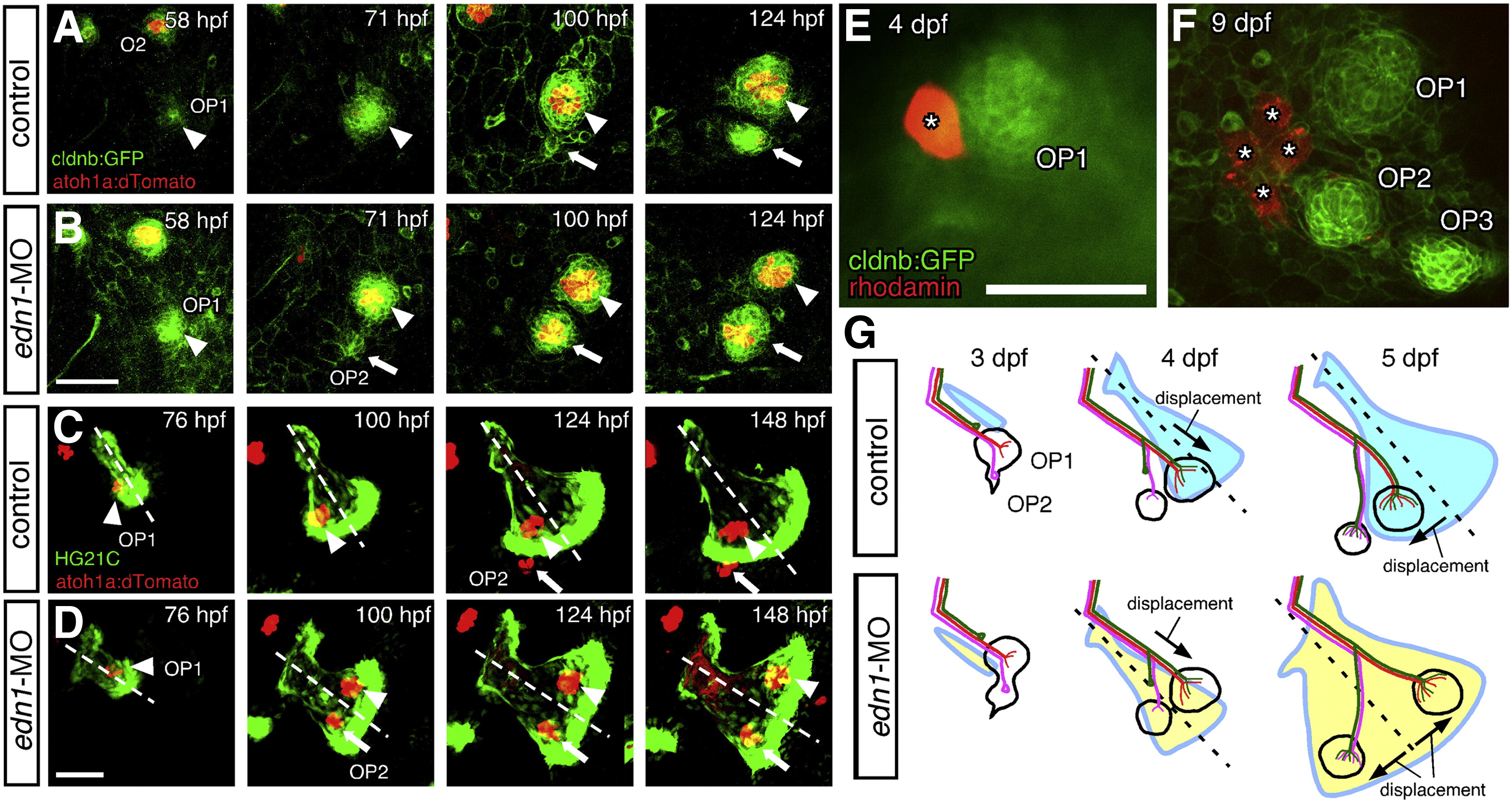
Fig. 6 Morphogenesis of the opercle correlates with neuromast patterning. (A–D) Time-course observations of control (A, C) and edn1-MO injected (B, D) embryos doubly transgenic for atoh1a:dtomato and cldnb:gfp (A, B), or HG21C (C, D). In morphant embryos, OP2 forms earlier than in control embryos (A, B). In control embryos, both OP1 and OP2 are located ventral to the opercle midline (C). In contrast, in morphant embryos, OP1 is located on the dorsal half of the opercle and moves away from OP2, which is located on the ventral opercle (D). Arrowheads and arrows indicate OP1 and OP2, respectively. (E, F) Single peridermal cell was labeled with rhodomine-dextrane and traced its fate, showing proliferation of peridermal cells relative to the neuromasts. (G) Schematic drawings of the developing opercular lateral line system in control and morphant embryo. The stage of the embryo is indicated in each panel. Scale bars: 50 μm.
Reprinted from Developmental Biology, 340(2), Wada, H., Ghysen, A., Satou, C., Higashijima, S.I., Kawakami, K., Hamaguchi, S., and Sakaizumi, M., Dermal morphogenesis controls lateral line patterning during postembryonic development of teleost fish, 583-594, Copyright (2010) with permission from Elsevier. Full text @ Dev. Biol.