
|
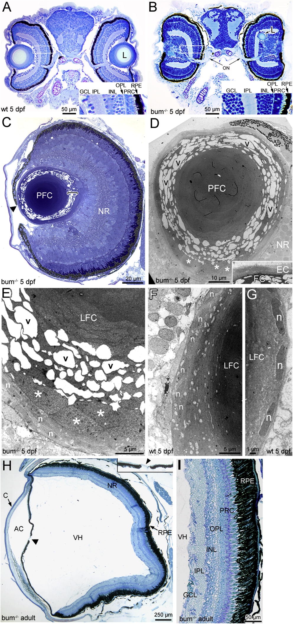
Fig. 4 The lens in 5 dpf bum-/- zebrafish shows a severe degeneration of secondary lens fibre cells and can be ectopically located in the neural retina. (A–F) Histology of the eyes in larval (A–G) and adult bum-/- zebrafish (H, I). (A, B) Toluidine blue-stained transversal sections through the central part of the eyes of 5 dpf sibling (wild-type, wt; A) and bum-/- (B) zebrafish larvae. Insets in bottom-right corners show magnifications of the boxed areas in the retinas of the eyes shown on the left in A and B, respectively. (C) Semi-thin section of a bum-/- eye at 5 dpf. The higher magnification of the lens reveals that in the outer lens cortex, the secondary lens fibre cells appear detached from each other with large empty spaces between them (see bracket indicating this region at the lens’ posterior pole). The central lens nucleus, however, seems to be composed of morphologically normal primary lens fibre cells. Note that part of the iris reaches beyond the centre of the lens (black arrowhead) leaving only a small, lopsided pupil opening. (D–G) Thin-section electron microscopy images of 5 dpf bum-/- (D, E) and wild-type lenses (F–G). The images of mutant lenses show that the initial stages of fibre cell differentiation occur normally with the cells (and their nuclei) adopting a flattened shape during cellular elongation. Soon after the onset of differentiation (3–4 cells into the lens), however, the differentiating secondary lens fibre cells appear to swell, increasing in thickness by a factor of up to ten (asterisks). A further 1–2 cells deeper into the lens, the secondary fibre cells appear to detach from each other and/or fragment. This results in large empty, vacuolar spaces (v) between fibre cell fragments. The fibre cells of the lens nucleus by contrast appear to have differentiated normally. Such swelling, detachment and fragmentation of secondary lens fibres is never observed in age-matched wild-type sibling lenses (F, G). (H, I) Morphology of the adult eye in bum-/- as revealed in transverse sections through the central part of the eye. Except for the conspicuous absence of the lens and an almost closed pupil opening (arrowhead and inset in H), the morphology of the eye in bum-/- mutant zebrafish is comparable to that of wild-type individuals, with a neural retina displaying all characteristic layers, a retinal pigment epithelium interdigitating with the well-developed photoreceptor outer segments, as well as a normally formed anterior chamber, vitreous humour cavity and cornea. A higher magnification of part of the retina of an adult bum-/- mutant zebrafish shows normal retinal layering (I). Abbreviations: AC, anterior chamber; C, cornea; EC, lens epithelial cells; GCL, ganglion cell layer; INL, inner nuclear layer; IPL, inner plexiform layer; L, lens; LFC, lens fibre cells; n, nuclei of differentiating secondary lens fibre cells; NR, neural retina; ON, optic nerve exiting the retina; OPL, outer plexiform layer; PFC, primary lens fibre cells; PRC, photoreceptor cells; RPE, retinal pigment epithelium; v, vacuolar spaces; VH, vitreous humour.
Reprinted from Mechanisms of Development, 127(3-4), Schonthaler, H.B., Franz-Odendaal, T.A., Hodel, C., Gehring, I., Geisler, R., Schwarz, H., Neuhauss, S.C., and Dahm, R., The zebrafish mutant bumper shows a hyperproliferation of lens epithelial cells and fibre cell degeneration leading to functional blindness, 203-219, Copyright (2010) with permission from Elsevier. Full text @ Mech. Dev.