
|
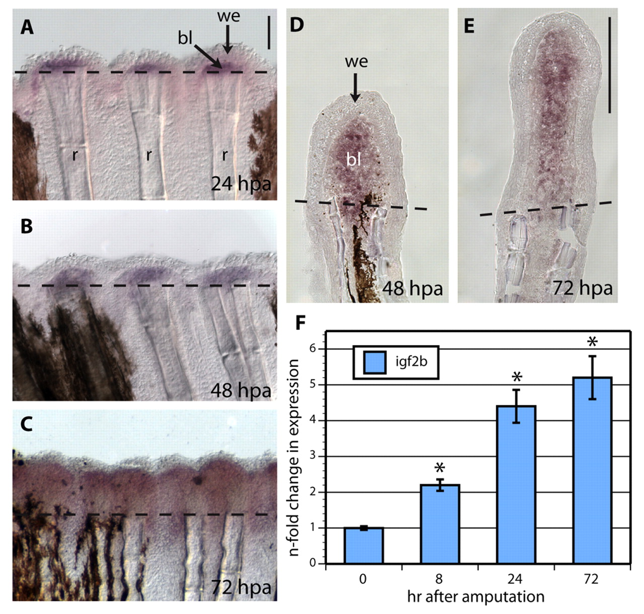
Fig. 1 igf2b is expressed in the blastema of regenerating fins. (A-C) Whole-mount in situ hybridization with igf2b mRNA antisense probe (purple) reveals gene expression at the distal region underneath the wound epidermis at 24 (A), 48 (B) and 72 (C) hpa. Dashed lines indicate the amputation plane. The regenerated tissue is located above the dashed lines. (D,E) In situ hybridization performed on cryosections at 48 and 72 hpa demonstrates igf2b expression in the blastema. (F) Quantitative RT-PCR determination of igf2b mRNA in regenerating fins at 8, 24 and 72 hpa relative to control fins at 0 hpa, which as calibrator samples were normalized to 1.00. Error bars represent s.e.m. *P<0.001; n=3 samples, each was prepared from 10-15 fins. bl, blastema; r, ray with skeletal elements; we, wound epidermis. Scale bars: 100 μm in A,E.