
|
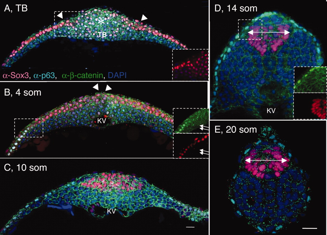
Fig. 1 Shaping of the posterior neural tube (PNT). A-E: Transverse sections through the tailbud region (at the level of Kupffer′s vesicle [KV]) of wild-type (WT) tailbud (TB) (A), 4 somites (som; B), 10 som (C), 14 som (D), and 20 som (E) embryos, labeled with α-Sox3C (neural tissue, pink), α-p63 (epidermis, nuclear labeling, turquoise), α-β-catenin (cell surface marker, green), and DAPI (4′,6-diamidine-2-phenylidole-dihydrochloride; nucleus, blue). Dorsal side is up in these and all subsequent images of transverse sections. Insets in (B,D) show separated channels in boxed areas; in insets p63-positive cells are labeled in green and Sox3C-positive cells are in red. The asterisk in A indicates the region of the tailbud expressing low levels of Sox3C. White arrowheads point to the medial edge of the neural domain. B: White arrows in insets point to cells coexpressing Sox3C and p63. Double arrows in (D,E) indicate the width of the neural tube. KV, Kupffer′s vesicle; som, somites; TB, tailbud. Scale bar = 20 μm.