
|
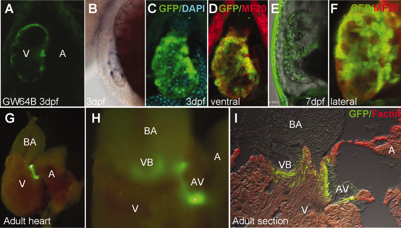
Fig. 5 Developmental changes in a CET line with early ventricle-specific expression (GW64B). A: EGFP expression at 3 dpf was found in the ventricle (ventral view). B: Same stage larvae stained by WISH using anti-gfp probe (lateral view). C, D, F, I: Double immunolabelling with MF20(red) performed on 3-dpf (C, D), 7-dpf (F), and adult heart section (I). C-F: The ventricular expression revealing trabeculated myocardium was maintained until 7 dpf. In the adult heart, EGFP expression was found at the A-V junction (G), and at the VB junction, where it defines valves (H, I). All images are GW64B. A, atrium; A-V, atrio-ventricular valve; BA, bulbus arteriosus; V, ventricle; VB, ventriculo-bulbar valve.