
|
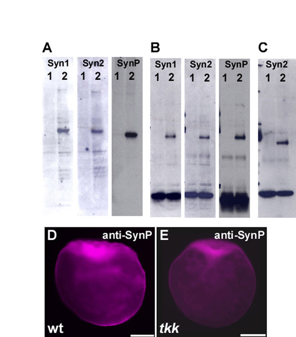
Fig. S8 Evaluation of anti-Syntabulin monoclonal and polyclonal antibodies. (A) Immunoblot detection of Syntabulin in lysates of HEK293T cells that were transfected with zebrafish Syntabulin expression plasmid (lane 2) or not transfected (lane 1). Ten microliters of 1 ml lysates of transfected HEK293T cells in a 6-cm dish were used. (B) Lysates from embryos just after fertilization (lane 2) or 60 mpf (lane1) were immunoprecipitated and immunoblotted with anti-Syntabulin antibodies. (C) Lysates from tkk (lane 1) or wild-type (lane 2) embryos just after fertilization were immunoprecipitated and immunoblotted with anti-Syntabulin (anti-Syn2) antibodies. (D,E) Immunostaining of wild-type and tkk embryos (fixed just after fertilization) with polyclonal anti-Syntabulin antibody (anti-SynP). Mouse monoclonal antibodies, anti-Syn1 and anti-Syn2, were raised against GST fusion proteins containing amino acids 14-94 and 102-271, respectively (see Fig. S2). Rabbit polyclonal antibody, anti-SynP, was raised against His-tagged protein containing amino acids 1-120 of Syntabulin. Anti-SynP was affinity purified with the antigen-conjugated column. Scale bars: 200 μm.