
|
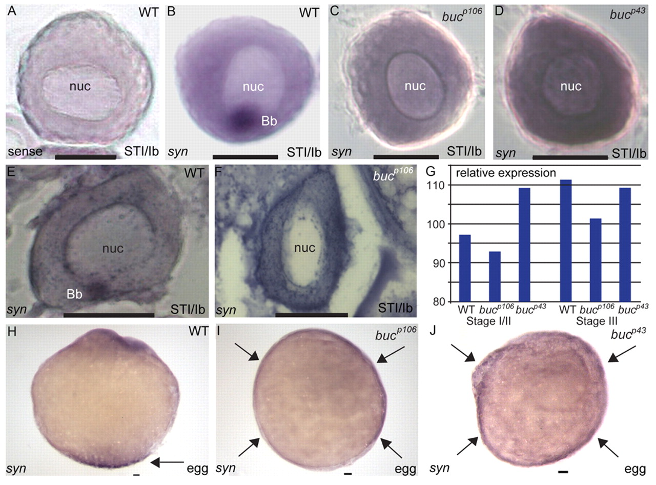
Fig. 6 Balbiani body-mediated localization of syntabulin at the vegetal pole. (A-D) Localization of syntabulin RNA in wild-type and bucky ball mutant (bucp106 and bucp43) stage Ib (STI/Ib) zebrafish oocytes. Staining by whole-mount in situ hybridization with sense (A, control) or antisense (B-D) riboprobes. In wild-type oocytes, syntabulin RNA localizes to the Balbiani body (A,B; 84%, n=104). In bucp106 (C; 97%, n=87 oocytes from three females) and bucp43 (D; 100%, n=36 from one female) mutant oocytes, syntabulin is not spatially restricted or localized. (E,F) Localization of syntabulin RNA. In wild-type oocytes (E), syntabulin localizes to the Balbiani body, whereas syntabulin is detected throughout the cytoplasm in bucp106 mutants (F; 100%, n=27 from one female). (G) Quantification of syntabulin expression in wild-type and buc oocytes. Although syntabulin is not localized in buc mutants, the transcripts are present in early and late stage oocytes. (H-J) Localization of syntabulin RNA in wild-type and buc activated eggs. In activated eggs from wild type, syntabulin shows polarized localization (H; 100% n=37 from two females). In activated eggs from bucp106 (I; 100%, n=44 from three females) and bucp43 (J; 100%, n=8 from one female) mutants, syntabulin is detected around the circumference. Arrows indicate localization of syntabulin at the vegetal pole in wild type and its circumferential expansion in buc eggs. nuc, nucleus; Bb, Balbiani body. Scale bars: 50 μm.