
|
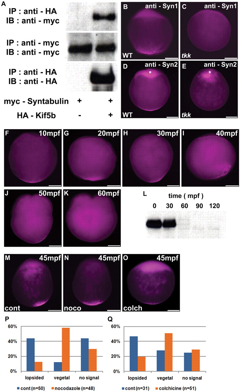
Fig. 5 Microtubule-dependent transportation of Syntabulin. (A) Interaction between Syntabulin and Kif5b. HEK293T cells were transfected with expression vectors of Myc-tagged Syntabulin and HA-tagged Kif5b. IP, immunoprecipitation; IB, immunoblotting. (B-E) Detection of Syntabulin with anti-Syntabulin monoclonal antibodies (anti-Syn1, anti-Syn2) in wild-type and tkk zebrafish embryos at early zygote stage (10 mpf). Expression of Syntabulin at the animal pole (asterisks in D,E) is non-specific as it is detected in tkk embryos, which lack significant amounts of Syntabulin (see Fig. S8C in the supplementary material). (F-K) Time course of Syntabulin localization in the wild type. Immunostaining with anti-Syn1. (L) Immunoblotting of anti-Syntabulin (anti-Syn2) immunoprecipitates. (M-O) Localization of Syntabulin at 45 mpf in wild-type embryos treated with 1 μg/ml nocodazole (N) or 0.4 mg/ml colchicine (O) just after fertilization, versus untreated control (M). (P,Q) Quantification of Syntabulin localization at 45 mpf showing lopsided or vegetal pole localization, or undetectable expression in embryos treated with nocodazole (P) or colchicine (Q), versus control (P,Q). Shown are lateral views with animal pole to the top. Scale bars: 200 μm.