
|
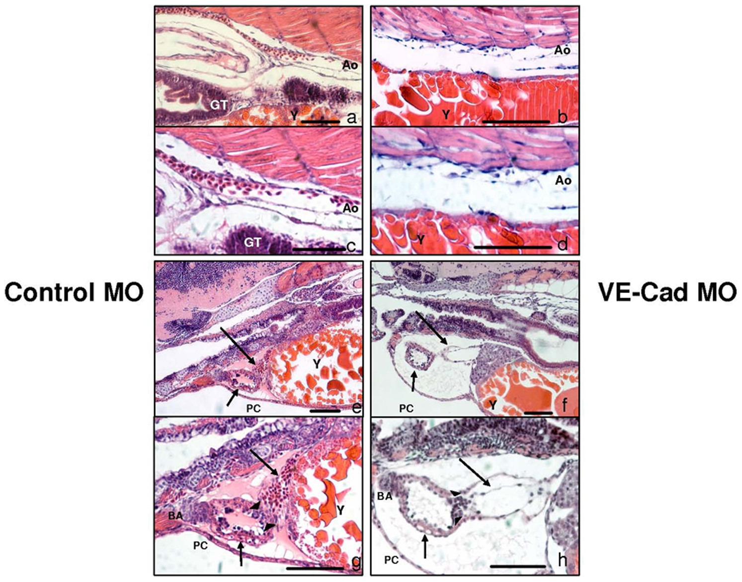
Fig. 5 VE-cadherin knockdown produces persistent, excessive endocardial/myocardial separation.
H+E staining at medium (300x) and high power (600x) aortic images of control (a,c) and VE-Cad knockdown (b,d) embryos at 96 hpf. The aorta of both control MO and VE-cad MO embryos appears contiguous. Medium (200x) and high power (400x) images of the thoracic region of control MO (e,g) and VE-cad MO (f,h) embryos. The atria of control embryos are intact (long arrows e,g), while those of knockdown embryos are elongated, with persistent endocardial detachment from the myocardial layer (long arrows f,h). The ventricles are intact in both control and knockdown embryos, though VE-cad MO embryos have thinner ventricular walls (short arrows e–h). Pericardial edema is present only in VE-cad MO embryos, however both types show a normal-appearing bulbus arteriosus, normal A–V valve formation (arrowheads) and blood within the aortic arches. Ao, Aorta; BA, Bulbus Arteriosus; GT, Gut Tube; PC, pericardium;Y, Yolk, Scale bars 75 μm.