
|
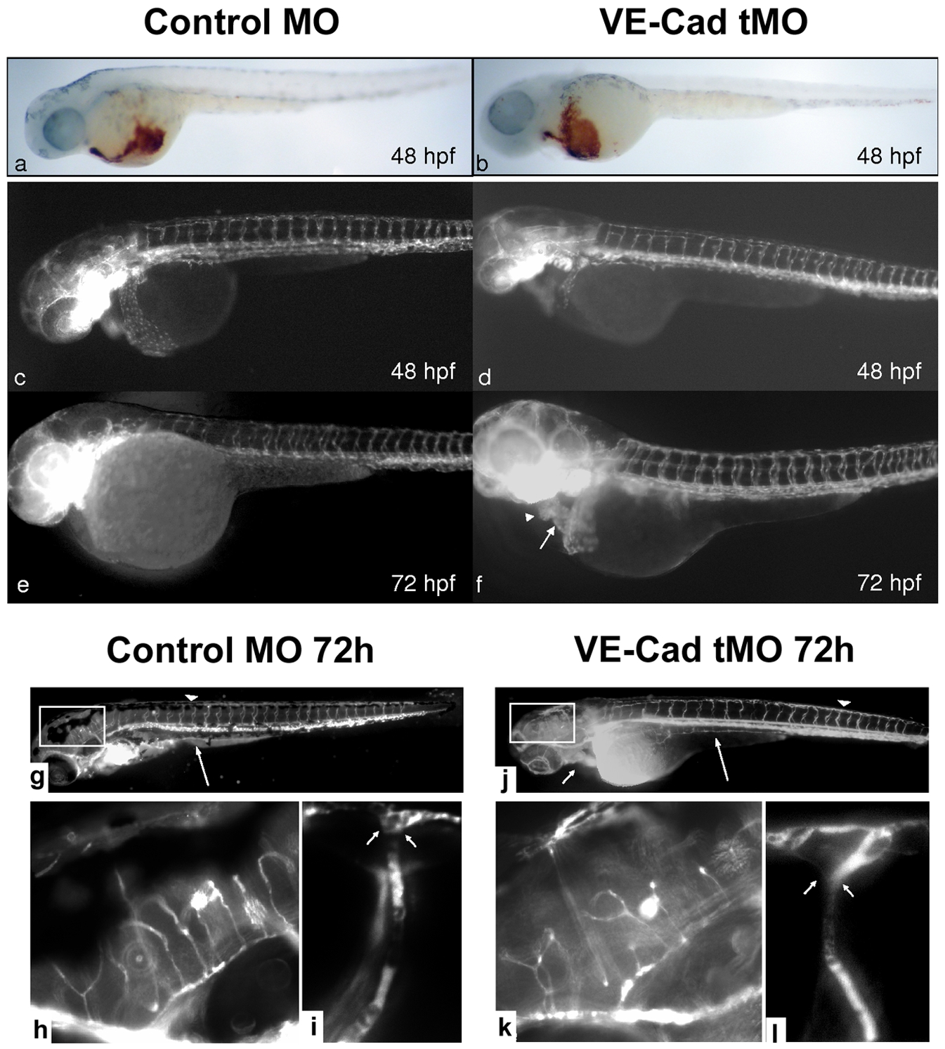
Fig. 3 Loss of VE-cadherin does not affect primitive hematopoiesis, vascular sprouting or peripheral vessel integrity.
Embryos at 48 hpf were stained with O-dianisidine for erythrocytes. No overall difference is observed between control (a) and Ve-cadherin knockdown embryos (b, 40x). Transgenic fli1:gfp embryos were injected with control MO and VE-cad MO and observed by epifluorescence microscopy. Vasculogenesis and intersomitic sprouting appears unaffected by VE-Cadherin knockdown (d) at 48 hpf compared to controls. At 48 and 72 hpf, despite normal vasculature, the endocardium of knockdown embryos (d,f) shows a linear atrial (arrow) and ventricular (arrowhead) orientation within a dilated pericardium (e). Low power images (g,j, 40x) of Control-MO and VE-Cad MO injected embryos show normal sprouting of the intersegmental vessels and DLAV (arrowheads) as well as the subintestinal vessels (long arrow) in both embryos. Impaired cardiac loopin,g is noted in VE-cad MO injected embryos (j, short arrow). Medium power images of the boxed regions (h,k 200x) demonstrate similar sprouting patterns between control and knockdown embryos in the head vasculature. Images of intersegmental/DLAV junctions (i,l 300x) show intact vessels and no extravasation in either control MO or VE-cad MO injected embryos (junctions at arrowheads I,l).