
|
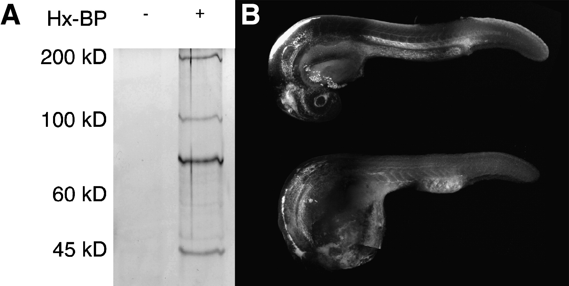
Fig. 6 Activity based protease labeling in zebrafish embryos. (A) Embryonic homogenates were mixed with the hydroxamate-based MMP probe (Hx-BP) carrying a biotin moiety, crosslinked by ultraviolet irradiation, and the biotinylated proteins collected by precipitation with streptavidin beads. The precipitated proteins were then resolved by sodium dodecyl sulfate–polyacrylamide gel electrophoresis and detected by silver staining. The left (blank) lane shows that no proteins from homogenates are bound nonspecifically by the beads, but in the right lane, strong bands at roughly 50, 72, 95, and 190kD are apparent, suggesting that Hx-BP is able to biotinylate proteins of these molecular weights. (B) Embryos were injected with Hx-BP carrying a rhodamine moiety, and then either crosslinked by ultraviolet irradiation (top), or left unirradiated (bottom) before fixation and examination by confocal microscopy. The crosslinked embryo shows strong rhodamine fluorescence in the ventricles of the brain, hatching gland, the mesenchymal regions of the head, surrounding the lens, myotome boundaries, the perichordal sheath, and the extracellular matrix separating the mesoderm from the ectoderm in the elongating tail. The unirradiated embryo shows strong fluorescence in the ventricles of the brain, and weak ubiquitous fluorescence elsewhere. This suggests that the Hx-BP probe is being specifically localized at sites where active MMPs are remodeling the extracellular matrix in the embryo.