
|
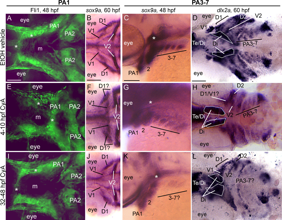
Fig. 9 Early and late Cyclopamine treatments lead to CNCC defects. Ventral (A,B,D,E,F,H,I,J,L) or lateral (C,G,K) views at 48 hpf (A,C,E,G,I,K) or 60 hpf (B,D,F,H,J,L) of vehicle-treated wild type (A-D) or Cya-treated wild type (E-L) embryos that are Fli1gfp positive (A,E,I) or labeled with RNA probe for sox9a (B,C,F,G,J,K) or dlx2a (D,H,L). (A,E,I) By 48 hpf, vehicle-treated wild type embryos (A) display proper patterning of anteriormost CNCC (asterisks), as do 32-48 hpf Cya-treated embryos (I); however, 4-10 hpf Cya-treated embryos (E) show mispatterning of anteriormost CNCC. (B,F,J) Vehicle treated embryos display normal sox9a expression in the dorsal (D1) and ventral (V1) CNCC condensations in the first arch (B), as do 32-48 hpf Cya-treated embryos (J). In 4-10 hpf Cya-treated embryos, ventral (V1) CNCC condensations maintain sox9a expression, while sox9a is greatly reduced in the dorsal (D1) region of the first arch. (C,D,G,H,K,L) Treating wild type embryos with vehicle or Cya from 4-10 hpf does not influence sox9a expression at 48 hpf(C,G) or dlx2a at 60 hpf (D,H) within posterior arch residing CNCC, while treating wild type embryos with Cya at 32-48 hpf leads to reduction in sox9a gene expression at 48 hpf (K) and dlx2a at 60 hpf (L) within CNCC in the posterior arches. Asterisks in (C,G,K) indicate sox9a expression in mesoderm-derived polar cartilages. Scale bar: 50 μM.