
|
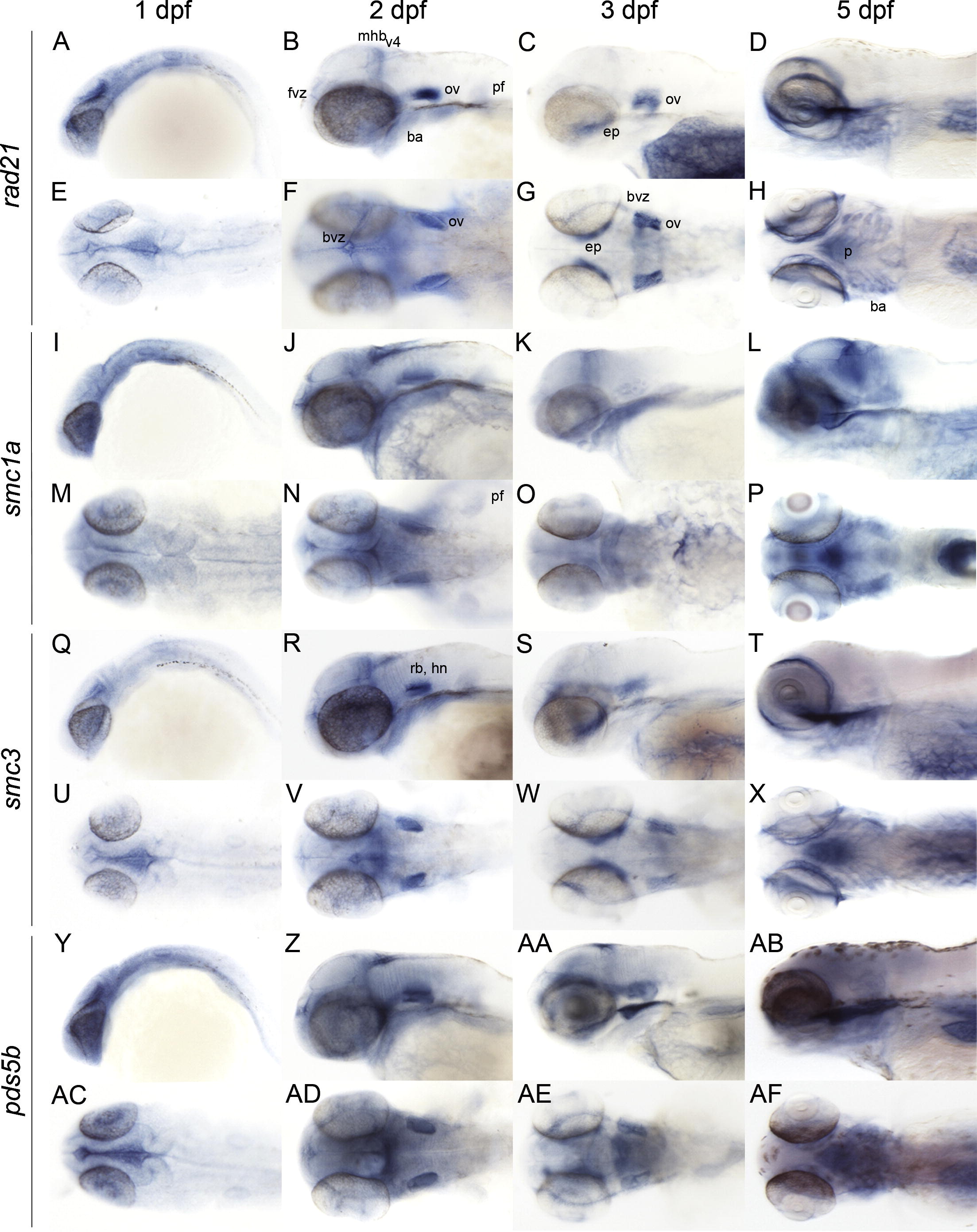
Fig. 2 Late embryonic and larval expression (1–5 dpf) of genes encoding zebrafish cohesin subunits. Whole mount in situ hybridization of zebrafish at the indicated stages with antisense riboprobes detecting the following mRNAs: (A–H) rad21. (I–P) smc1a. (Q–X) smc3. (Y–AF) pds5b. A–D, I–L, Q–T, Y–AB: lateral views. E–H, M–P, U–X, AC–AF: dorsal views. Anterior is to the left for all. ba, branchial arches; bvz, brain ventricular zone; ep, ethmoid plate; fvz, forebrain ventricular zone; hn, hindbrain neurons; mhb, midbrain hindbrain boundary; ov, otic vesicle; p, pharynx; pf, pectoral fin; rb, rhombomeres boundaries; v4, 4th ventricle.
Reprinted from Gene expression patterns : GEP, 9(8), Mönnich, M., Banks, S., Eccles, M., Dickinson, E., and Horsfield, J., Expression of cohesin and condensin genes during zebrafish development supports a non-proliferative role for cohesin, 586-594, Copyright (2009) with permission from Elsevier. Full text @ Gene Expr. Patterns