
|
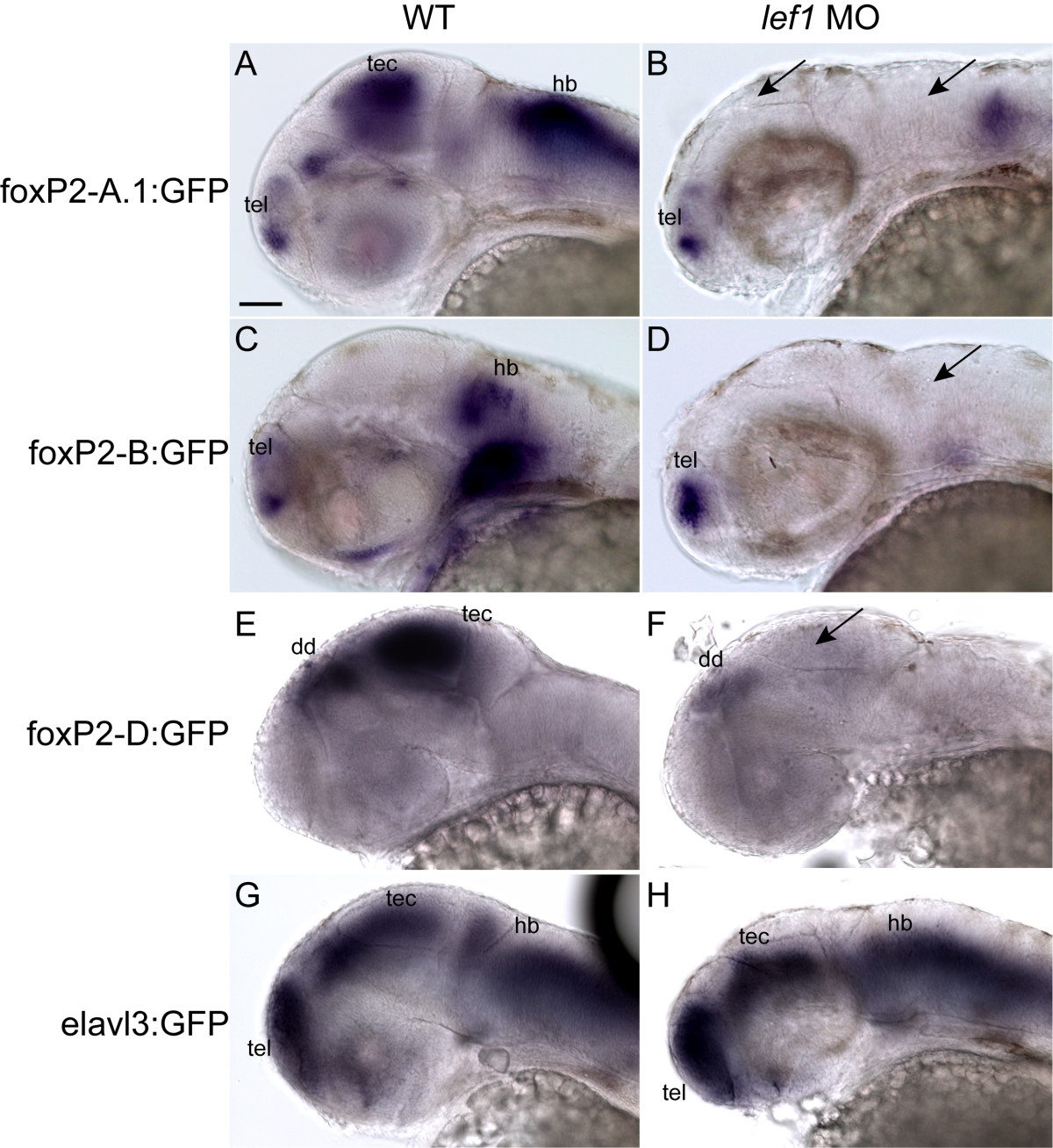
Fig. 6 lef1 is necessary for expression from foxP2-enhancerA.1, foxP2-enhancerB, and foxP2-enhancerD. Whole-mount gfp in situs at 36 hpf; anterior to left, dorsal up, eyes removed. Scale bar = 50 μm. Conditions (wild type, wt; or lef1 morphant, lef1 MO) are shown above the panels, enhancer names to left. (Abbreviations: dd, dorsal diencephalon; hb, hindbrain; tec, tectum; tel, telencephalon.) (A) Tg(foxP2-enhancerA.1:EGFP)zc44 expresses in telencephalon, tectum, and hindbrain. (B) Tg(foxP2-enhancerA.1:EGFP)zc44 embryo injected with lef1 morpholino lacks GFP expression in tectum and hindbrain (arrows), although telencephalic expression persists. (C) Tg(foxP2-enhancerB:EGFP)zc41 expresses in telencephalon and hindbrain. (D) Tg(foxP2-enhancerB:EGFP)zc41 embryo injected with lef1 morpholino lacks GFP expression in the hindbrain (arrow), but telencephalic expression is present. (E) Tg(foxP2-enhancerD:EGFP)zc47 expresses in dorsal diencephalon and tectum. (F) Tg(foxP2-enhancerD:EGFP)zc47 embryo injected with lef1 morpholino lacks GFP expression in the tectum (arrow), but dorsal diencephalic expression persists. (G) Tg(elavl3:EGFP)zf8 embryo shows GFP expression in all post-mitotic neurons. (H) Tg(elavl3:EGFP)zf8 embryo injected with lef1 morpholino still has GFP expression in the tectum and hindbrain.