
Image
|
Figure Caption
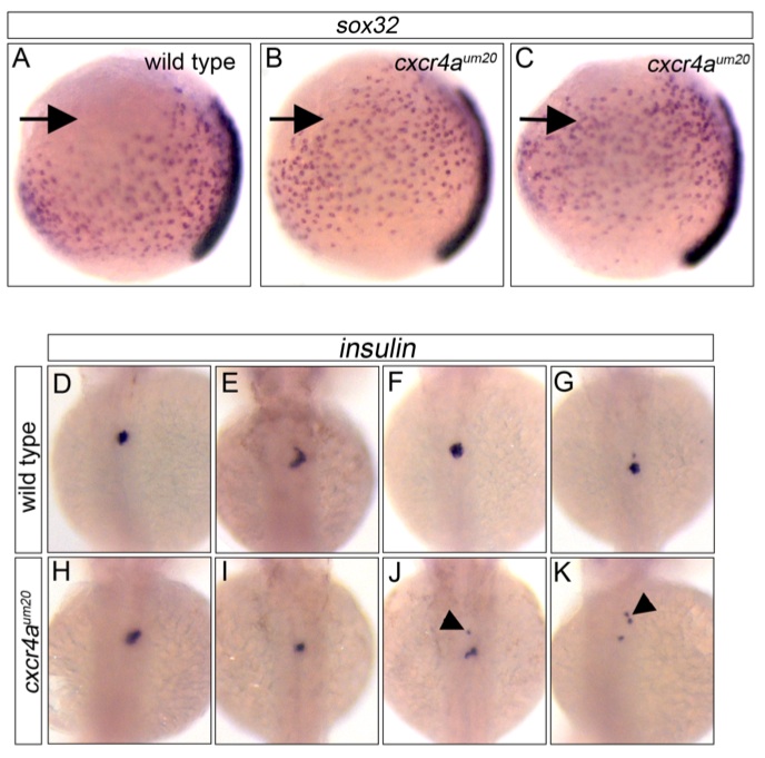
Fig. S6 Endoderm patterning defects in cxcr4a mutant embryos. A-C, sox32 in situ hybridization of 90% epiboly embryos. Dorsal is to the right, animal pole up. Note anterior displacement of endodermal cells in cxcr4a um20 mutant embryos (B, C, arrows) when compared to wild type embryo (A). D-K, insulin in situ hybridization at 55 hours post fertilization in 4 representative wild type embryos (D-G), compared to cxcr4a um20 mutant embryos (H-K). Note displaced insulin expressing cells (arrowheads) in J, K.
Figure Data
Acknowledgments
This image is the copyrighted work of the attributed author or publisher, and
ZFIN has permission only to display this image to its users.
Additional permissions should be obtained from the applicable author or publisher of the image.
Full text @ Genes & Dev.