
|
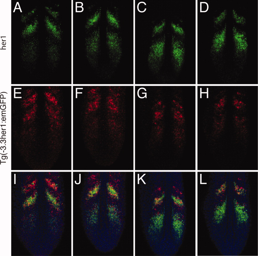
Fig. 5 The -3.3-kb enhancer does not accurately recapitulate her1 expression in the anterior presomitic mesoderm (PSM). Double fluorescent in situ hybridization of four Tg(-3.3her1:emGFP) embryos. GFP, green fluorescent protein. Three images of a single confocal section are shown for each embryo: (A,E,I), (B,F,J), (C,G,K), and (D,H,L), respectively. A-D: Expression of her1. Embryos are arranged to indicate progression of waves of her1 transcription through the somitogenesis cycle. E-H: Expression of emGFP mRNA. I-L: Merged images showing expression of her1 (green) and emGFP (red). Nuclei stained with propidium iodide are colored blue.