
|
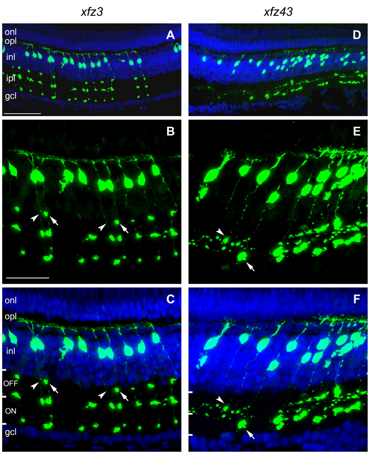
Fig. 5 Axon terminal patterns within the adult retina define distinct types of bipolar cells. (A-C) Confocal images of a retinal cryosection, labelled with an anti-GFP antibody (green) and counterstained with DAPI (blue), from an adult Tg(Gal4-VP16;UAS:eGFP)xfz3 fish (6 months). Higher magnification images of the middle part in (A) are shown in (B, C). The image in (B) is the same as in (C) but without DAPI. Arrows in (B, C) indicate a bouton-like axon terminal structure in the OFF layer, which is present in one bipolar cell type but absent in the other (arrowheads). (D-F) Retinal cryosection from an adult Tg(Gal4-VP16;UAS:eGFP)xfz43 fish. The confocal images are arranged in the same way as shown for the other line. Arrows in (E, F) indicate a large bouton-like structure in the ON sublamina, which is present at the axon terminal of one bipolar cell type. A second type of bipolar cells shows terminal arborisation within the OFF sublamina (arrowheads). ON and OFF sublamina of the inner plexiform layer are indicated by bars in (C, F). Scale bars: 50 μm (A, D); 25 μm (B, C, E, F). Abbreviations: gcl, ganglion cell layer; inl, inner nuclear layer; ipl, inner plexiform layer; onl, outer nuclear layer; opl, outer plexiform layer.