
Image
|
Figure Caption
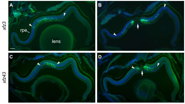
Fig. 4 Regional distribution patterns of eGFP labelled bipolar cells in adult retina. Fluorescence images of retinal cross-sections from adult fish (6 months) from the transgenic lines Tg(Gal4-VP16;UAS:eGFP)xfz3 (A, B), and Tg(Gal4-VP16;UAS:eGFP)xfz43 (C, D). Sections were stained with an anti-GFP antibody (green) and counterstained with DAPI (blue). All images are oriented with the ventral side to the left. Arrowheads mark the central region containing eGFP labelled bipolar cells. The optic nerve is indicated by arrows (B, D). Scale bar: 100 μm. Abbreviation: rpe, retinal pigmented epithelium.
Figure Data
Acknowledgments
This image is the copyrighted work of the attributed author or publisher, and
ZFIN has permission only to display this image to its users.
Additional permissions should be obtained from the applicable author or publisher of the image.
Full text @ BMC Neurosci.