
|
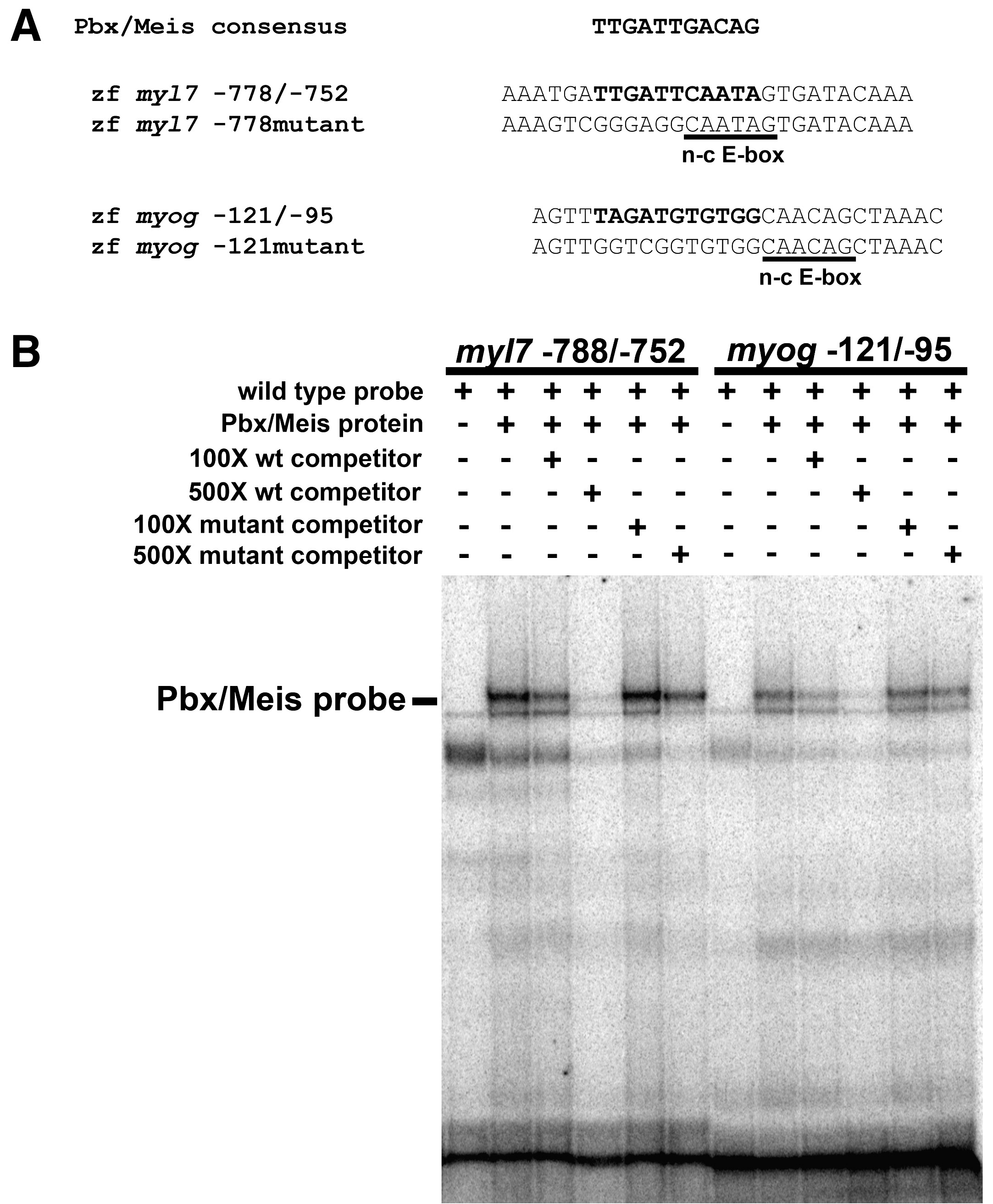
Fig. 6 Pbx proteins bind the myl7 promoter in vitro. (A) Pbx/Meis consensus binding site sequence (Berkes et al., 2004) aligned relative to putative Pbx binding site sequences (highlighted in bold) in the zebrafish myl7 promoter and myogenin (myog) promoter. The non-canonical E-box (n-c E-box) sites are underlined. Also shown are sequences of mutated Pbx binding sites. Sequences represent oligos used in EMSA. (B) Pbx4 and Meis3 proteins were generated by in vitro translation using a reticulocyte lysate and subjected to EMSA with the specified myl7 or myog probes. Pbx/Meis-bound probe band is indicated. The non-specific bands were observed in multiple gel shifts. Addition of unlabeled myl7 -778/-752 probe or myog -121/-95 probe decreases binding of Pbx/Meis, whereas addition of mutant unlabeled probes does not.
Reprinted from Developmental Biology, 333(2), Maves, L., Tyler, A., Moens, C.B., and Tapscott, S.J., Pbx acts with Hand2 in early myocardial differentiation, 409-418, Copyright (2009) with permission from Elsevier. Full text @ Dev. Biol.