
Image
|
Figure Caption
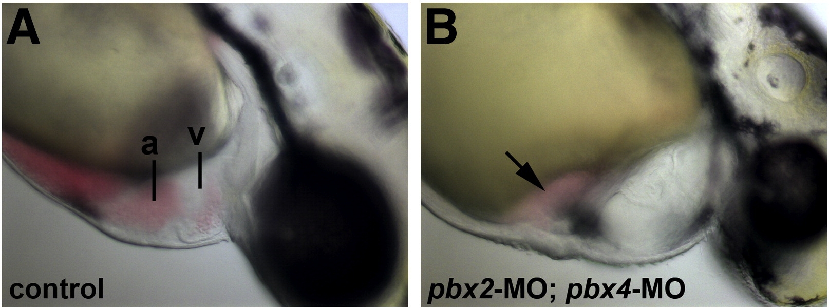
Fig. 1 Defective heart development in pbx2-MO; pbx4-MO embryos. (A–B) Lateral views of live (A) wild-type and (B) pbx2-MO; pbx4-MO embryos at 48 hpf. pbx2-MO; pbx4-MO embryos show mild pericardial edema and blood pooled near the atrium (arrow). Anterior is to the right. a, atrium. v, ventricle.
Figure Data
Acknowledgments
This image is the copyrighted work of the attributed author or publisher, and
ZFIN has permission only to display this image to its users.
Additional permissions should be obtained from the applicable author or publisher of the image.
Reprinted from Developmental Biology, 333(2), Maves, L., Tyler, A., Moens, C.B., and Tapscott, S.J., Pbx acts with Hand2 in early myocardial differentiation, 409-418, Copyright (2009) with permission from Elsevier. Full text @ Dev. Biol.