
Fig. 6
- ID
- ZDB-IMAGE-090904-35
- Publication
- Bollig et al., 2009 - A highly conserved retinoic acid responsive element controls wt1a expression in the zebrafish pronephros
- All Figures
- Figures for Bollig et al., 2009
|
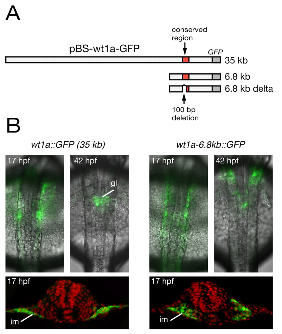
Fig. 6 The conserved region upstream of wt1a is required for its expression in the intermediate mesoderm. (A) Schematic representation of wt1a reporter constructs. The upper row represents the plasmid pBS-wt1a-GFP, which was used for the generation of wt1a::GFP transgenic zebrafish. In addition, two truncated wt1a-GFP plasmids are shown containing a 6.8 kb genomic fragment (middle row) and the same fragment lacking 100 bp within the conserved region (lower row). A detailed illustration of the deleted 100 bp region is shown in Fig. 5B. (B) Overlay of brightfield transmission and fluorescence images from wt1a::GFP (left) and wt1a-6.8kb::GFP (right) transgenic embryos at 17 and 42 hpf (top). For detailed analysis, embryos at 17 hpf were stained with anti-GFP antibody (green) and were sectioned (bottom). Counterstaining with DAPI is shown in false color (red). gl, glomeruli; im, intermediate mesoderm.