
Fig. 3
- ID
- ZDB-IMAGE-090904-33
- Publication
- Bollig et al., 2009 - A highly conserved retinoic acid responsive element controls wt1a expression in the zebrafish pronephros
- All Figures
- Figures for Bollig et al., 2009
|
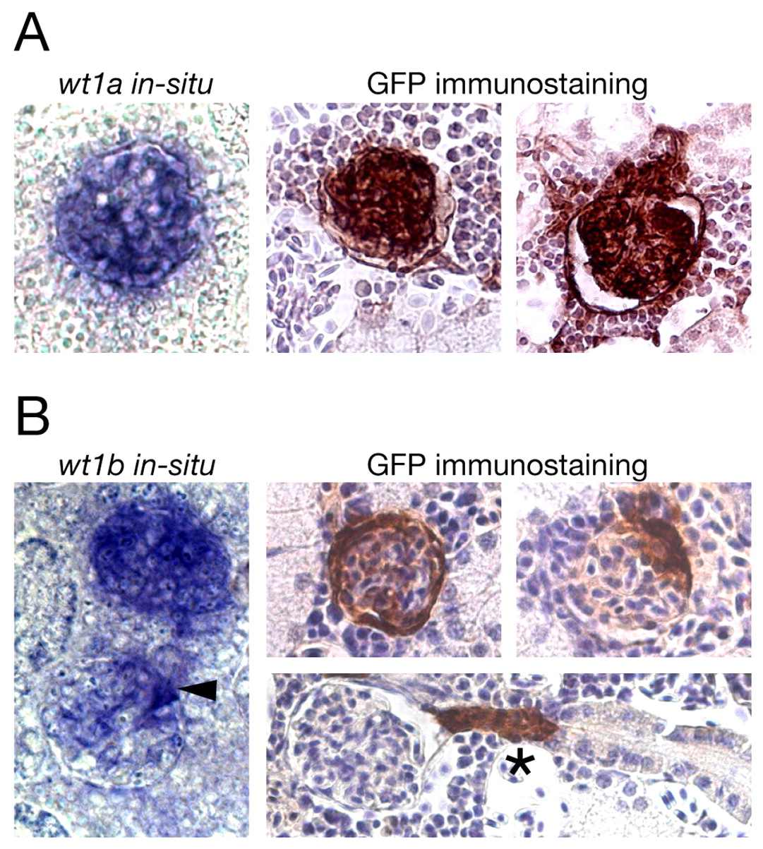
Fig. 3 GFP expression in the mesonephros of adult transgenic zebrafish recapitulates expression of wt1 paralogs. (A,B) In situ hybridization for wt1a (A) and wt1b (B) on sections of wild-type mesonephros (left) and GFP immunostainings on sections of wt1a::GFP line 1 (A) and wt1b::GFP line 1 (B) mesonephros (right) are shown. The kidneys were taken from 4- to 6-month old wild-type and transgenic zebrafish. In the immunostainings, cell nuclei are stained blue (Hematoxylin counterstaining) and GFP-positive cells are brown. Arrowhead marks a glomerulus in which only a subset of cells is labeled, asterisk denotes a GFP-positive neck region.