
|
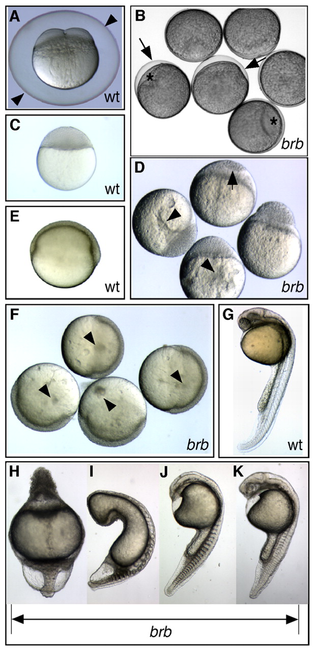
Fig. 1 brom bones mutants display impaired egg activation and dorsoventral patterning defects. (A) Wild-type (wt) 4-cell-stage zebrafish embryo at 60 mpa, with a fully elevated chorion (arrowheads). (B) Failed chorion elevation (arrows) and cytoplasmic segregation in brom bones mutant (brb) eggs at 60 mpa. Mutants have small blastodiscs (*) and slightly opaque yolk. (C) A wild-type embryo at oblong stage (a midblastula stage). (D) Moderately affected mutant embryos exhibit incomplete cellularization (arrow) and display large cytoplasmic inclusions in the yolk (arrowheads) at oblong stage. (E,F) Small cytoplasmic inclusions (arrowheads) are visible in the yolk of mildly affected mutant embryos (F) at gastrula stages, but not in wild-type embryos (E). (G) Wild-type embryo at 1 dpf. (H) Radially ventralized brom bones mutant embryo and (I-K) brom bones mutant embryos with decreasing strength of ventralization at 1 dpf. G-K are dissecting microscope images; rostral is towards the top and dorsal is to the right.