
|
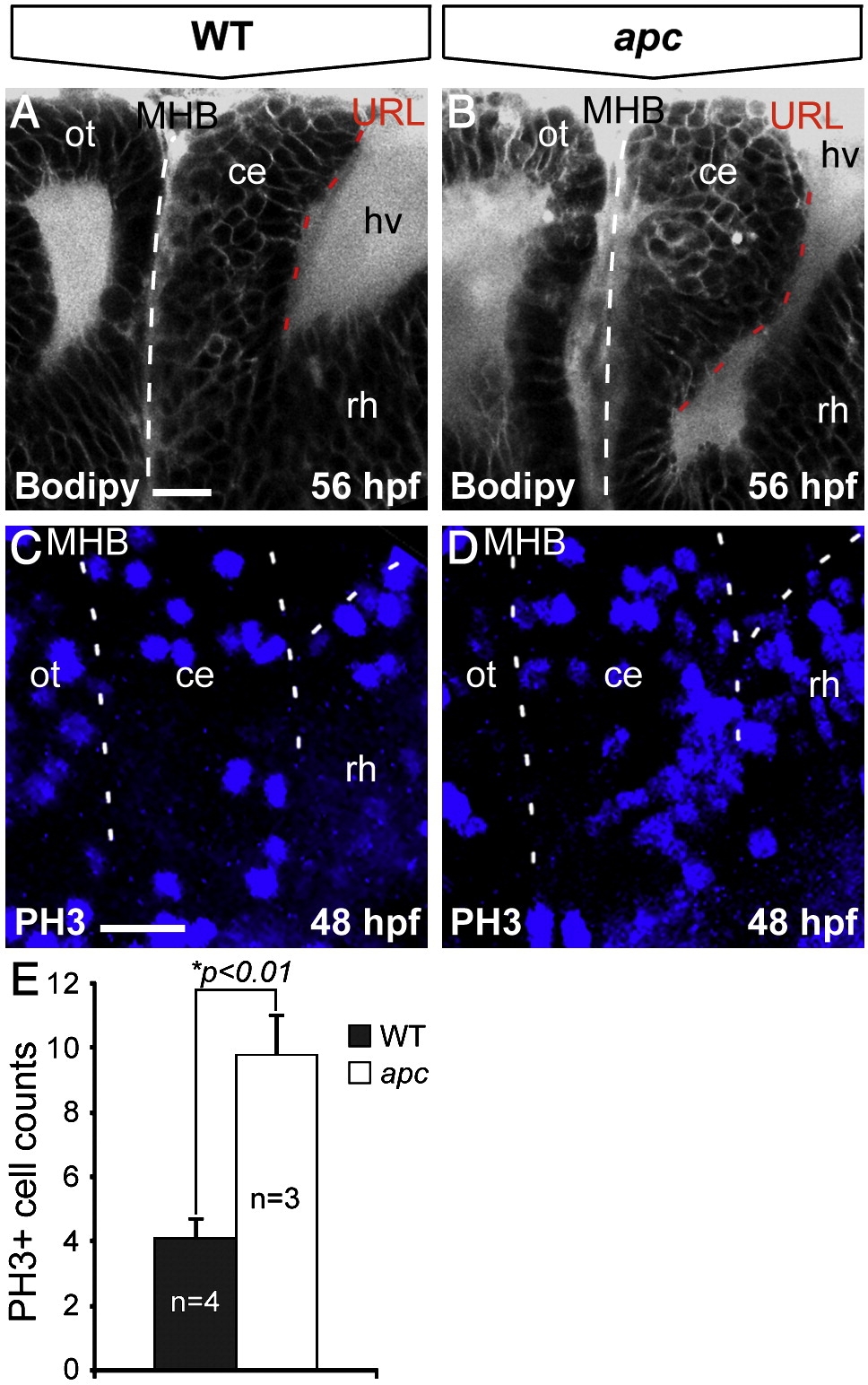
Fig. 4 Loss of Apc function results in unscheduled proliferation in the cerebellum. Lateral view, anterior to the left. (A, B) Bodipy vital staining marking cell membranes reveals that the apc cerebellum (B) is enlarged and curves towards the MHB (indicated with dashed line) as compared to wild-type cerebellum (A). (C, D) Phosphohistone-3 (PH3) immunolabeling that marks mitotic cells identifies unscheduled increased proliferation in the mutant cerebellum (D) as compared to wild-types (A). Left dashed line indicates MHB, bifurcating dashed lines on the right indicate boundary between cerebellum and the hindbrain/rhombomere 1. (E) The mutant cerebellum shows a significantly higher number of PH3+ cells as determined by Student′s t-test (*p < 0.01). Error bars represent standard error of the mean (SEM). ce, cerebellum; hv, hindbrain ventricle; MHB, mid–hindbrain boundary; ot, optic tectum; rh, rhombomere1. Scale bar 15 μm.
Reprinted from Developmental Biology, 331(2), Paridaen, J.T., Danesin, C., Elas, A.T., van de Water, S., Houart, C., and Zivkovic, D., Apc1 is required for maintenance of local brain organizers and dorsal midbrain survival, 101-112, Copyright (2009) with permission from Elsevier. Full text @ Dev. Biol.