
|
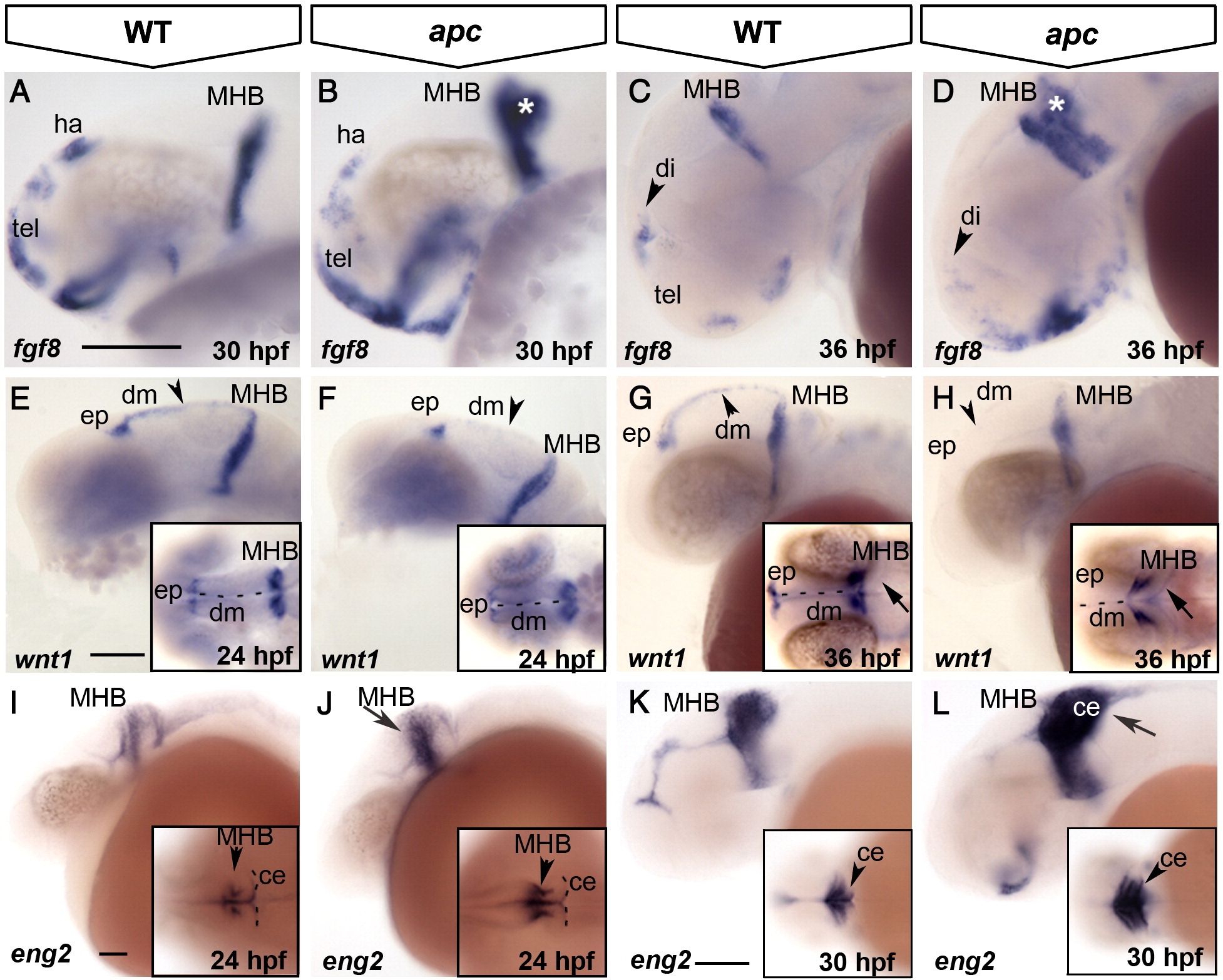
Fig. 3 Loss of Apc function compromises the IsO. Lateral view, anterior to the left. (A–D) fgf8 expression expands in the ventral telencephalon and into the cerebellum (asterisk) in apc mutants at 30 hpf (B). At 36 hpf (C, D), fgf8 is reduced in the habenula (arrowhead in panel D). (E–H) At 24 hpf, wnt1 expression is reduced in apc mutants (F) from the epiphysis and dorsal midline (arrowhead in panel F) and entirely absent from the dorsal midline at 36 hpf (H). wnt1 is weakly expanded into the mutant cerebellum (arrow in inset panel H). Inset - dorsal view. Dashed line indicates the midline. (I–L) At 24 hpf, eng2a expression is enhanced at the apc MHB (arrow and arrowhead in panel J; compare to inset in panel I). At 30 hpf (K, L), eng2a is expanded into the cerebellum (arrow in panel L and arrowhead in inset). Inset - dorsal view. Dashed line indicates cerebellum. ce, cerebellum; dm, dorsal midline; ep, epiphysis; ha, habenula; MHB, mid–hindbrain boundary; tel, telencephalon. Scale bar 125 μm.
Reprinted from Developmental Biology, 331(2), Paridaen, J.T., Danesin, C., Elas, A.T., van de Water, S., Houart, C., and Zivkovic, D., Apc1 is required for maintenance of local brain organizers and dorsal midbrain survival, 101-112, Copyright (2009) with permission from Elsevier. Full text @ Dev. Biol.