
|
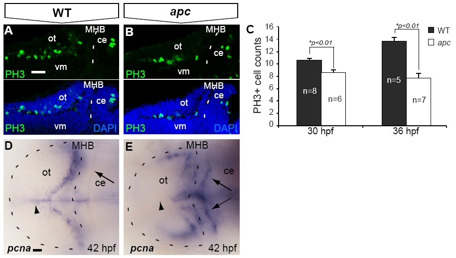
Fig. S5 Reduced proliferation in the tectum opticum of apc mutants. (A–C) At 30 hpf, confocal analysis identified fewer PH3+ mitotic cells in the apc mutant OT. (B) as compared to wild-types (A). (C) The reduction of PH3+ cell numbers was augmented during further development. (D, E) Reduction of proliferation in the mutant OT is corroborated by the absence of pcna labeling from the mutant OT midline (arrowhead; n = 5). Of note is enhanced ectopic expression of pcna in the mutant cerebellum (arrows). Statistics: two-tailed Student′s t-test, *p < 0.01. Error bars represent SEM. ce, cerebellum; MHB, mid–hindbrain boundary; ot, optic tectum; vm, ventral midbrain. Scale bar 100 μm.
Reprinted from Developmental Biology, 331(2), Paridaen, J.T., Danesin, C., Elas, A.T., van de Water, S., Houart, C., and Zivkovic, D., Apc1 is required for maintenance of local brain organizers and dorsal midbrain survival, 101-112, Copyright (2009) with permission from Elsevier. Full text @ Dev. Biol.