
|
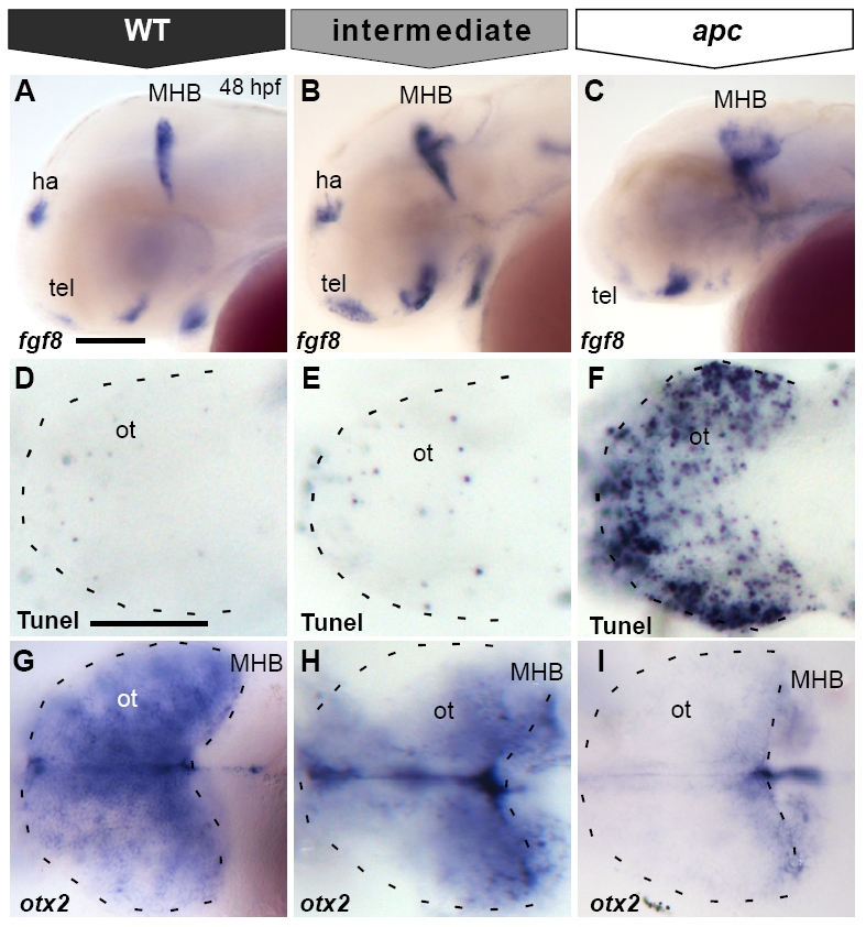
Fig. S3 Misexpression of apc-GFP mRNA in apc mutants results in various degrees of phenotypic rescue. (A–C) Lateral view. (D–I) Dorsal view, with eyes removed in (G–I). Anterior to the left. All embryos at 48 hpf. Phenotypes of fgf8 expression (A–C), apoptosis (D–F) and otx2 expression (G–I) as categorized in Figs. 6A and 8H, I. Wild-type or completely rescued apc-GFP injected mutant embryos show normal expression domains of fgf8 at the MHB, habenula (ha) and telencephalon (A), virtually no apoptosis in the tectum (D) and otx2 expression throughout the tectum (G). In partially rescued embryos, fgf8 expression is restored at the habenula and telencephalon, but is still expanded at the MHB (B), similarly to control mutants (C). Partially rescued mutants show a much improved cell survival as compared to the apc phenotype (F). However, there is still a low level of apoptosis in the tectum (E) as compared to the wild-type phenotype (D). otx2 expression is still slightly reduced in partially rescued mutants (H). ha, habenula; MHB, mid–hindbrain boundary; ot, optic tectum; tel, telencephalon. Scale bar 125 μm.
Reprinted from Developmental Biology, 331(2), Paridaen, J.T., Danesin, C., Elas, A.T., van de Water, S., Houart, C., and Zivkovic, D., Apc1 is required for maintenance of local brain organizers and dorsal midbrain survival, 101-112, Copyright (2009) with permission from Elsevier. Full text @ Dev. Biol.