
|
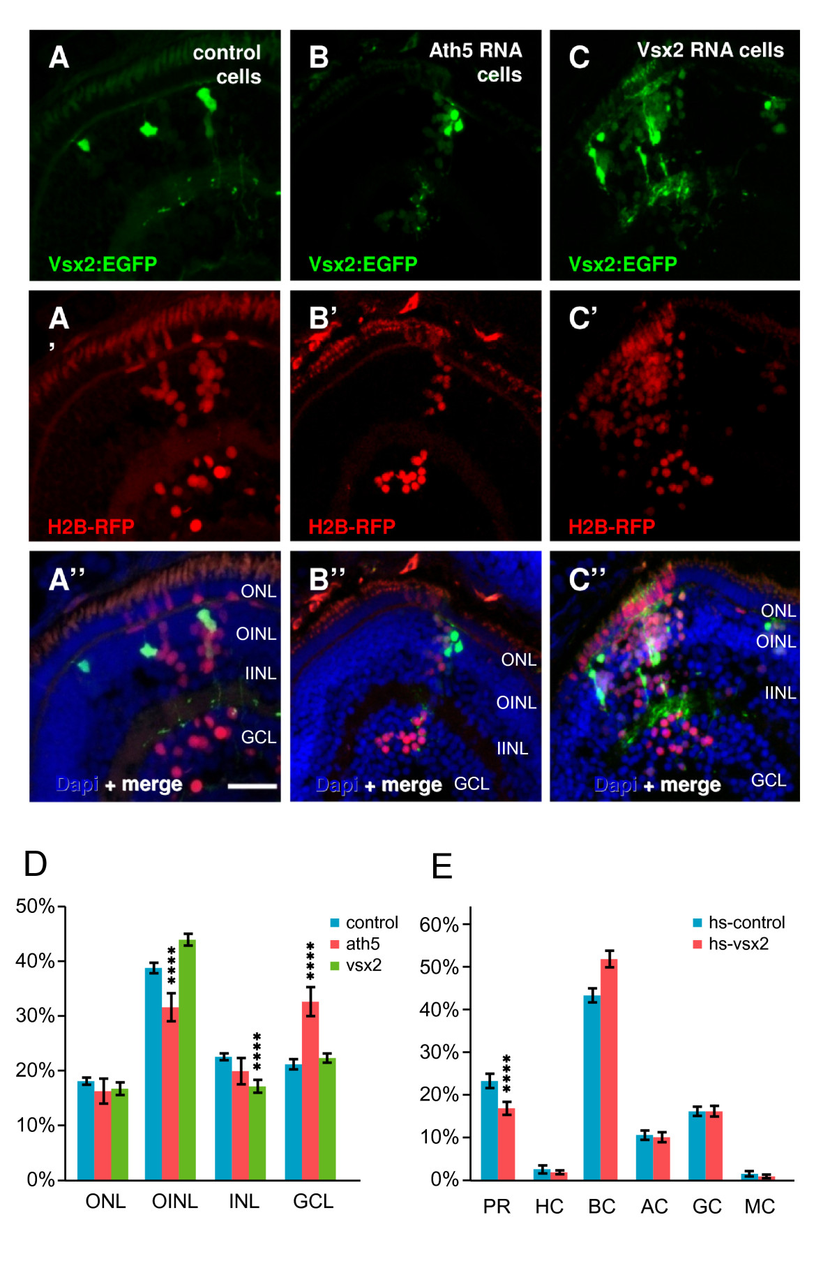
Fig. 14 Transplantation of wild type, Ath5, and Vsx2 overexpressing cells in wild-type embryos. (A-C)Tg(vsx2:GFP) transplanted into wild-type embryos after injection with H2B-RFP RNA (to mark all transplanted cells) and various constructs for overexpression. (A) Green-fluorescent protein (GFP)-expressing cells from control Tg(vsx2:GFP) become bipolar and Müller cells, whilst all (H2B-RFP-positive) cells become all kinds of cells in the expected frequency. (B) When co-injecting H2B-RFP RNA with ath5, the ganglion cell fate is promoted (more H2B-RFP cells in the GCL), but Vsx2:GFP cells still become bipolar and Müller cells. (C) Co-injection of H2B-RFP RNA with vsx2 promotes bipolar cell fate, seen by (D) a highly significant (p < 0.001) increase of H2B-RFP in the INL bipolar layer at the expense of amacrine cells (asterisks). (E) Overexpression using a heatshock vsx2 construct similarly results in an increased frequency of bipolar cells, this time at the expense of photoreceptors. Error bars indicate Standard Error of the Mean. AC, amacrine cell; BC, bipolar cell; GCL, ganglion cell layer; H, horizontal cell; hs, heat shock; IINL, inner half of inner nuclear layer; MC, Müller cell; OINL, outer half of inner nuclear layer; ONL, outer nuclear layer; PH, photoreceptor; RGC, retinal ganglion cell. Scale bar (A-C) 25 μm.