
|
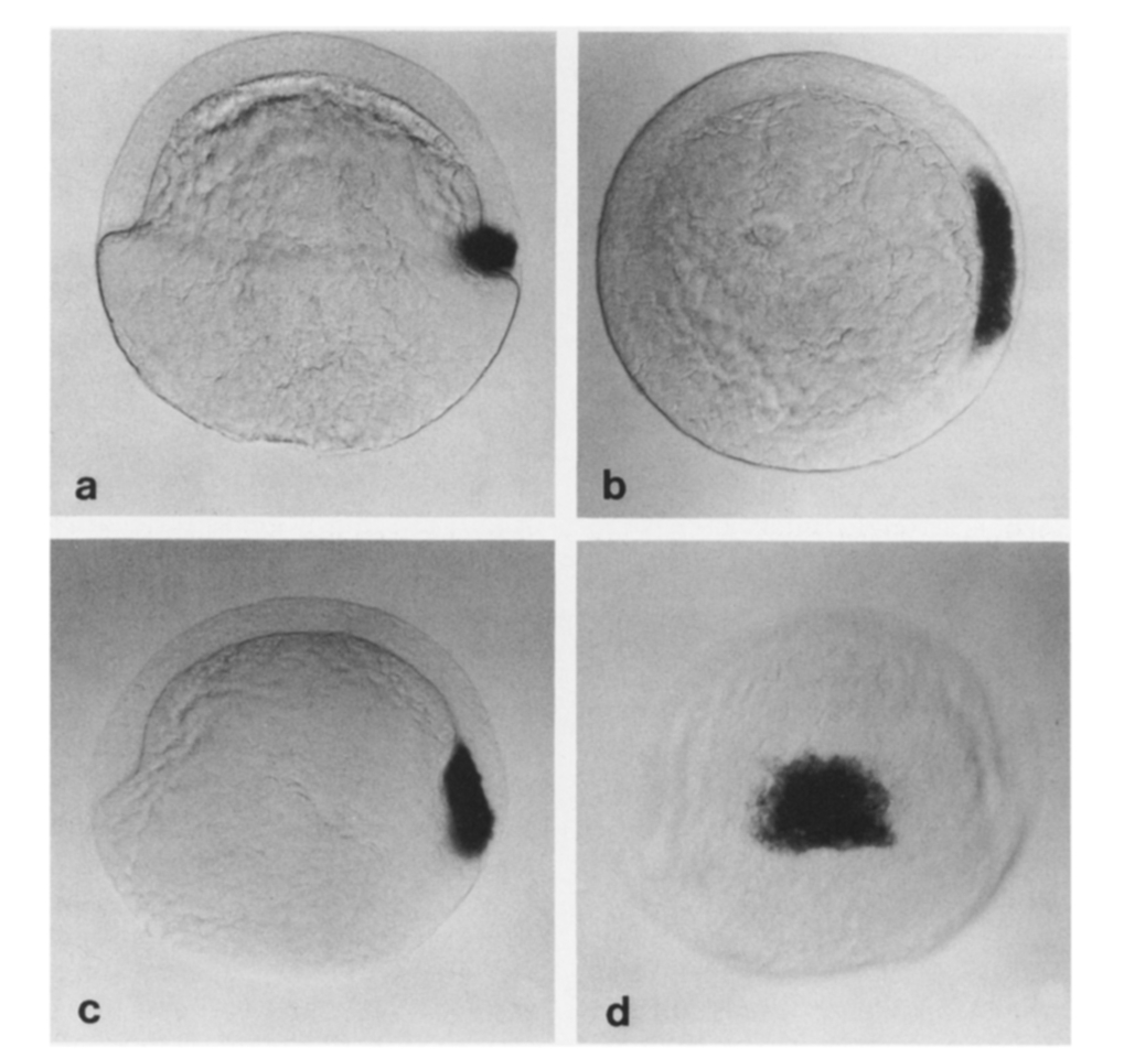
Fig. 1 Localization of gsc transcripts by in situ hybridization in wild-type late blastula and early gastrula. (a) Lateral view of an embryo at 45% epiboly (late blastula) showing gsc transcripts restricted to a group of deep cells at the dorsal margin of the embryo (animal pole up; dorsal, right). (b) Animal pole view of the same embryo. The territory of goosecoid expression occupies about 60° of the circumference of the embryo at the dorsal margin (dorsal, right; ventral, left). (e) Lateral view of an embryo at 60% epiboly (early gastrula). All goosecoid-expressing cells have involuted and the territory has begun to extend toward the animal pole. (d) Dorsal view of the same embryo showing that the gsc-expression territory now occupies the central part of the embryonic shield, the precursor of the prechordal plate (animal pole, up).
Reprinted from Developmental Biology, 164, Thisse, C., Thisse, B., Halpern, M.E., and Postlethwait, J.H., Goosecoid expression in neurectoderm and mesendoderm is disrupted in zebrafish cyclops gastrulas, 420-429, Copyright (1994) with permission from Elsevier. Full text @ Dev. Biol.