
|
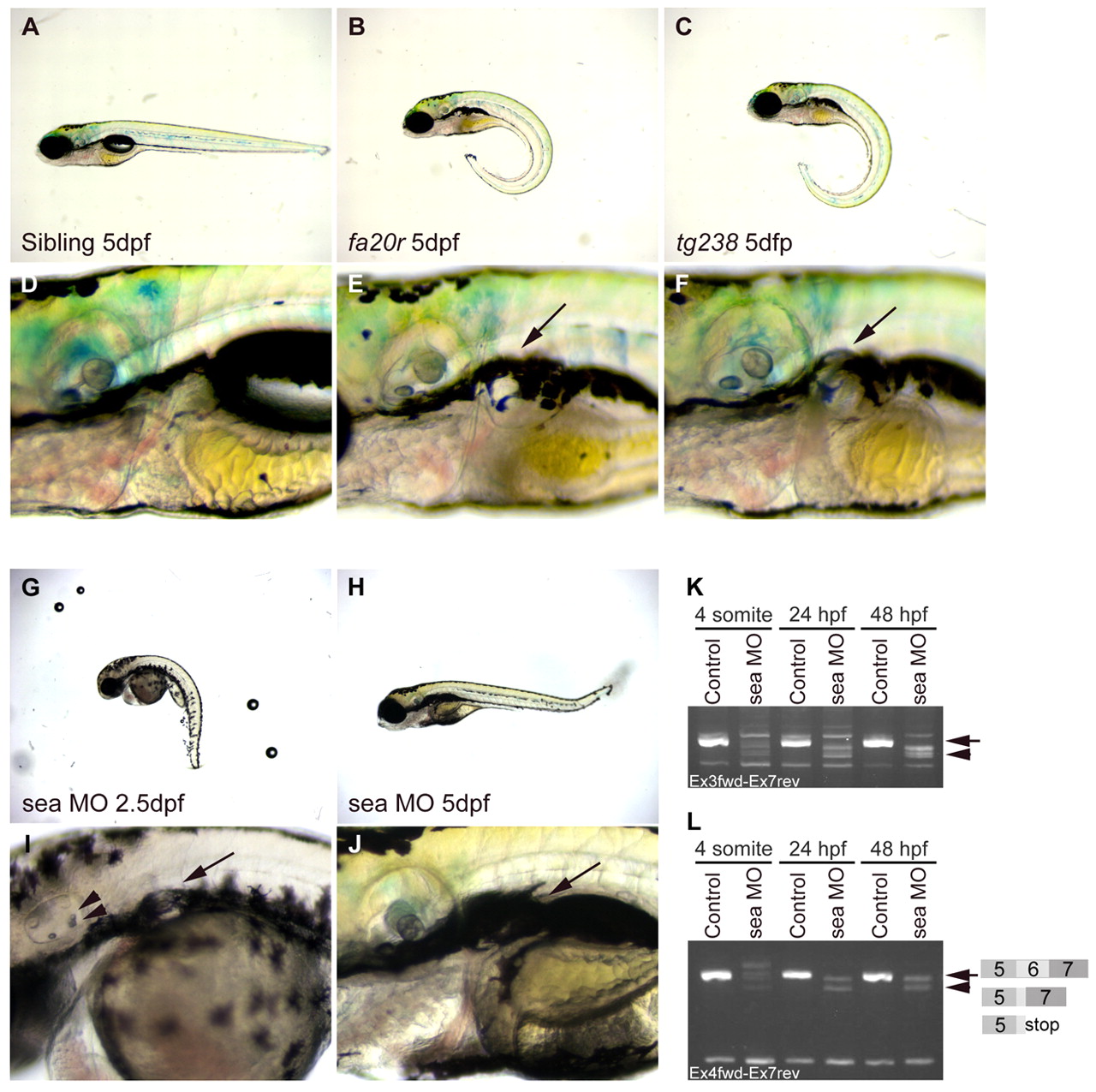
Fig. 2 seahorse mutants and sea MO-injected embryos displayed curly tail down and pronephric cyst phenotypes. (A-F) CTD and pronephric cyst phenotypes in sea mutants. (A) Siblings, (B) seafa20r and (C) seatg238a; mutant embryos have CTD phenotypes at 5 dpf. (D) Siblings, (E) seafa20r and (F) seatg238a embryos at higher magnification to show pronephric cysts at 5 dpf (arrows). (G-J) CTD and pronephric cyst phenotypes in sea MO-injected embryos. MO-injected embryos can recover from the CTD (H) and cyst phenotypes (J) by 5 dpf. Defects in otic vesicle and otolith formation (arrowheads) were observed in some sea MO-injected embryos (I). (K,L) RT-PCR from uninjected and sea MO-injected cDNA libraries at four somites, 24 hpf and 48 hpf. (K) Primers between exon 3 and exon 7 amplified a wild-type sea band at 635 base pairs (bp; arrow) and incorrectly spliced message in sea MO-injected embryos (arrowhead). (L) Primers between exon 4 and exon 7 amplified a wild-type sea band at 517 bp (arrow; exon 5-7) and incorrectly spliced message in sea MO-injected embryos (arrowhead). Incorrect splicing generated two main splice forms creating either a deletion and/or stop codon in exon 6 (indicated by the diagram in L).