
|
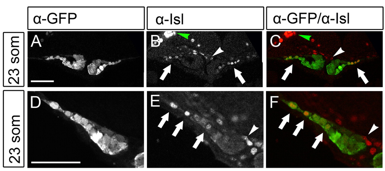
Fig. 3 Isl localization in cardiomyocytes of the future atrium. (A-F) Isl is expressed in cardiac cells located at lateral positions in the cardiac disk. Single z-scans of confocal images of Tg(cmlc2:eGFP) embryos at 23 somites immunofluorescently stained with α-eGFP and α-Isl antibodies. C and F show an overlay with eGFP in green and Isl in red. Images in D-F are magnified views of one side of the cardiac disk. Arrows indicate the eGFPposIslpos cells located at lateral positions (future atrium) of the cardiac disk. The white arrowhead indicates Isl-positive cells in the endocardium and the green arrowhead indicates the Isl signal in the trigeminal sensory ganglion. Scale bars: 50 μm.