
|
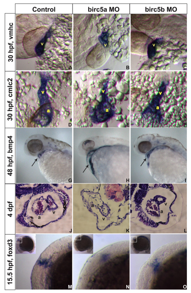
Fig. 5 Birc5 in cardiogenesis. In situ hybridizations (A-I, M-O) and histologic sections (J-L) on AB embryos. A-C: At 30 hpf, expression of cardiac ventricle marker vmhc is reduced with Birc5a depletion (A, B), and to lesser extent in Birc5b morphants (C). D-F: cmlc2 staining shows Birc5a (E) or Birc5b (F) morphants with impaired development of atrium and ventricle, compared to controls (D). G-I: At 48 hpf, bmp4 normally localizes in heart to reveal a ring-like structure, representing endocardial cushions of the atrio-ventricular valve (G, arrow). With depletion of Birc5a, bmp4 staining remains diffuse and ring structure is absent (H, arrow). In Birc5b-morphants, bmp4 localizes normally (I, arrow), but the ring is smaller. J-L: H&E stained histologic sections of hearts of normal embryos (J), and those depleted of Birc5a (K) and Birc5b (L) at 4 dpf. Birc5a morphants have thin-walled heart chambers, and little evidence of a-v valve formation. Birc5b-depleted embryos have smaller ventricles. M-O: Premigratory cardiac neural crest cells contributing to heart development, were detected by staining embryos at 15.5 hpf (13 somites) with foxd3. Compared to controls (M), premigratory neural crest cells were barely detectable in embryos depleted of Birc5a (N), and reduced in Birc5b morphants (O). a:atrium, v:ventricle.