
|
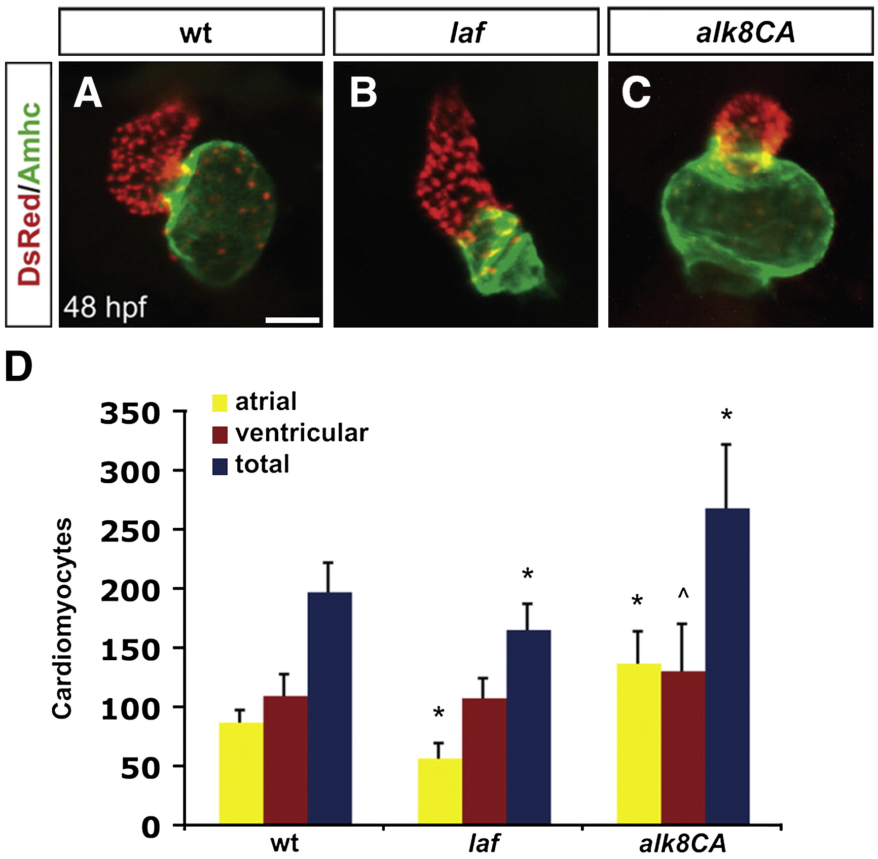
Fig. 1 Alk8 function regulates chamber proportionality. (A–C) Frontal views of hearts from wild-type (A), laf mutant (B) and alk8CA mRNA-injected (C) embryos at 48 h.p.f. Scale bar represents 50 μm. Immunofluorescence detects DsRed (red) in all cardiomyocyte nuclei and atrial myosin heavy chain (Amhc; green) in atrial cells. (A) In a wild-type heart, the ventricle (red) and atrium (green) exhibit typical looping and expansion. (B) In a laf mutant heart, the chambers are unlooped and dysmorphic. (C) In alk8CA mRNA-injected embryos displaying a v2–v3 class of ventralized body morphology (Kishimoto et al., 1997), the hearts are enlarged with a particularly striking expansion of the atrium. (D) Quantification of cardiomyocyte number in wild-type, laf mutant and alk8CA mRNA-injected embryos at 48 h.p.f. Graph indicates the mean and standard deviation for each data set; see also Table S1. Statistically significant differences from wild-type are indicated by asterisks (p < 0.005) or carets (p < 0.05). n = 29 for wild-type, n = 19 for laf mutants, and n = 10 for alk8CA mRNA-injected embryos. laf mutants and alk8CA mRNA-injected embryos have disproportionately small or large atrial chambers, respectively.
Reprinted from Developmental Biology, 328(2), Marques, S.R., and Yelon, D., Differential requirement for BMP signaling in atrial and ventricular lineages establishes cardiac chamber proportionality, 472-482, Copyright (2009) with permission from Elsevier. Full text @ Dev. Biol.