Image


                        Figure Caption


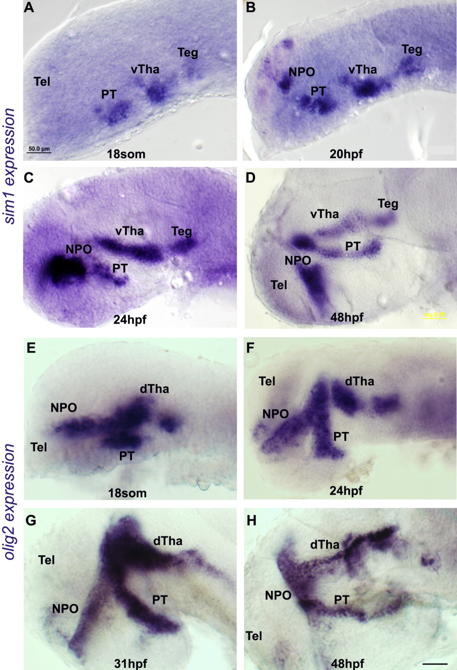
Fig. 1 Expression of sim1 and olig2 at different developmental stages. Micrographs of zebrafish embryos stained by whole mount in situ hybridization procedure using (A-D) Sim1 RNA probe and (E-H) Olig2 RNA probe. The different developmental stages are indicated. All images presented in lateral view, anterior to the left. dTha, dorsal thalamus; NPO, neurosecretory preoptic area; PT, posterior tuberculum; Teg, tegmentum; Tel, telencephalon; vTha, ventral thalamus. Scale bar = 50 μm.
                
                    
                        Figure Data


                        Acknowledgments


This image is the copyrighted work of the attributed author or publisher, and
ZFIN has permission only to display this image to its users.
Additional permissions should be obtained from the applicable author or publisher of the image.
      Full text @ Dev. Dyn.


        