
|
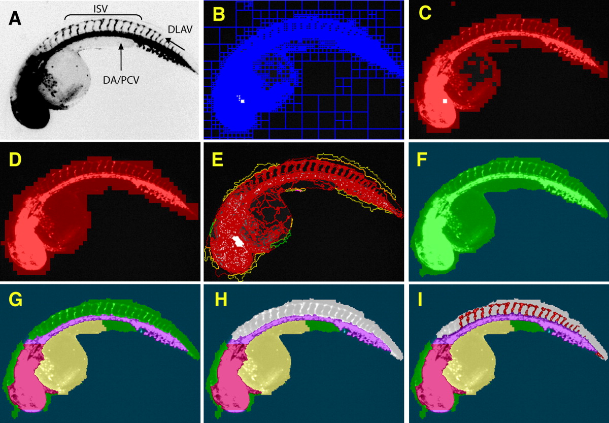
Fig. 1 Successive assembly of a hierarchical network identifies intersegmental vessels (ISV) in Tg(fli1:EGFP)y1 embryos. At 48 hours postfertilization (hpf), Tg(fli1:EGFP)y1 zebrafish embryos were imaged on an ArrayScan II high-content reader equipped with a x1.25 objective. A,B: The original image (A) was segmented based on pixel intensities and regional variability (B). C,D: Regions of high variability (C) were fused and expanded to provide a general outline (D). E,F: The outline was refined (E) to demarcate the whole zebrafish embryo (F). G,H: Specific subdomains within the embryo such as head, dorsal aorta/posterior cardinal vein, yolk (G), and dorsal area (H, gray) were then assigned through successive interactive loops of locally specific segmentation and classification, resulting in a hierarchical structure of the entire embryo with its subdomains. I: Knowledge generated during each prior classification step enabled the ruleset to specifically quantify ISV in the dorsal tail (red) without interference from other fluorescent regions. DA, Dorsal aorta; PCV, posterior cardinal vein; ISV, intersegmental vessels; DLAV, dorsal longitudinal anastomotic vessel.