
Fig. 2
- ID
- ZDB-IMAGE-090306-21
- Genes
- Publication
- Löhr et al., 2009 - Zebrafish diencephalic A11-related dopaminergic neurons share a conserved transcriptional network with neuroendocrine cell lineages
- All Figures
- Figures for Löhr et al., 2009
|
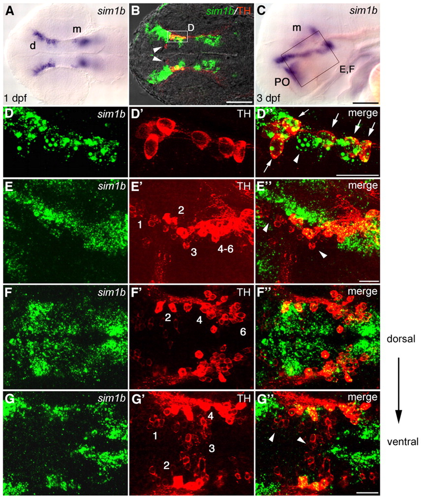
Fig. 2 sim1b is expressed in a subset of DA neurons in the ventral diencephalon. (A-G″) Expression of sim1b (A,C) and co-expression of sim1b (fluorescent whole-mount in situ hybridization, green) and TH (B,D-G″; immunohistochemistry red). (A,B) 1 dpf. (C) sim1b expression at 3 dpf. (D-D″) Higher magnification of area framed in B. All TH-positive cells in this region co-express sim1b (arrows), whereas some sim1b-expressing cells are negative for TH (arrowheads). (E-E″) Higher magnification of area framed in C. DA neuron groups 2, 4-6 colocalize with sim1b; DA neuron groups 1 and 3 do not co-express sim1b (arrowheads). (F-G″) Dorsal views at 3 dpf in more dorsal (F-F″) and more ventral (G-G″) coronal optical sections. (A,B,F-G″) Dorsal views, (C-E″) lateral views, anterior towards the left. Scale bars: in C, 100 μm for A,C; in B, 100 μm; in D″,E″,G″, 50 μm for D-G″. Images in B,D-G″ are z-projections of multiple adjacent focal planes, depths (B,D) 12 μm, (E) 30 μm and (F,G) 20 μm. d, diencephalon; m, mesencephalon; PO, preoptic region.