
|
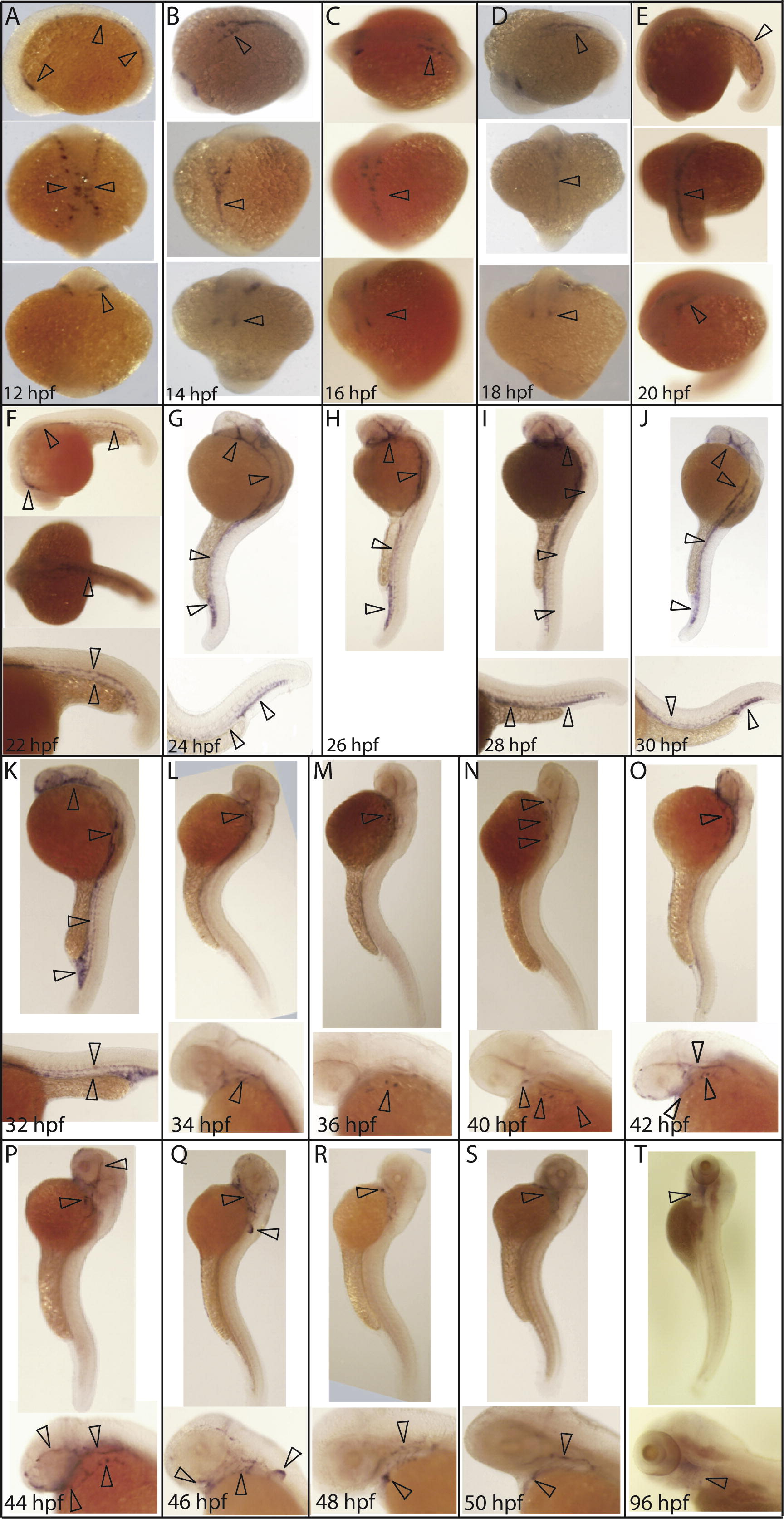
Fig. S1 Detailed time-course of erg expression during vasculogenesis. (A) erg expression was first observed at 12 hpf in mesodermal angioblasts. Later, erg positive cells are located in the midline and begin to coalesce into major axial and cranial vessels (B–E). (F) By 22 hpf, erg positive cells distinctly define the anterior lateral dorsal aortas, mandibular arches and ventral aorta; lateral dorsal aorta and posterior cardinal vein and posterior caudal artery and caudal vein. (G–J) Between 24 and 30 hpf, erg expression also expands to cranial primitive internal carotid artery, primordial midbrain channel, middle cerebral vein, anterior cerebral vein, and caudal and cranial divisions of the internal carotid artery. At 32 hpf (K), erg expression is retained in developing cranial, lateral and posterior vessels while faint expression is observed in developing intersegmental vessels. At 34 hpf (L), erg expression is lost in all major vessels, with expression then restricted to vessels in the developing pharyngeal arch region until 42 hpf (N). At 42 hpf (O), some expression is also seen in developing cranial vessels, although this is mostly lost by 46 hpf (Q), at which point expression is most prominent in the developing fin bud. By 50 hpf (S), expression is restricted to the primary head sinus, hepatic portal/common cardinal venous plexus, primitive internal carotid artery and mandibular arches. erg expression is still detectable in the aortic arches at 96 hpf (T). All panels are whole-mount in situ hybridisation analyses. Orientations are as follows. (A–E) Top panel: lateral; central panel dorsal; lower panel ventral. (F) Top panel: lateral; central panel dorsal; lower panel tail at higher magnification. (G–K) Top panel lateral, lower panel tail at higher magnification. (L–T) Top panel: lateral view, lower panel: head at higher magnification.
Reprinted from Mechanisms of Development, 126(3-4), Ellett, F., Kile, B.T., and Lieschke, G.J., The role of the ETS factor erg in zebrafish vasculogenesis, 220-229, Copyright (2009) with permission from Elsevier. Full text @ Mech. Dev.