
|
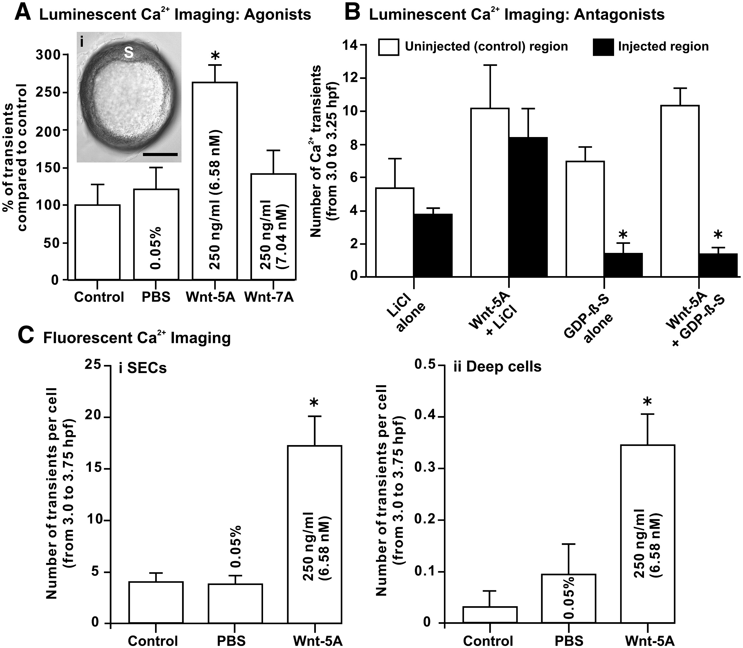
Fig. 9 The effect of agonists and antagonists of the Wnt/Ca2+ signaling pathway on the SEC Ca2+ transients generated during the dorsal-biased Ca2+ signaling window. Ca2+ transient data collected via luminescent f-aequorin (A and B) and fluorescent calcium green-1 dextran (C and D) imaging. (A) Comparison of the number of DCW SEC-generated Ca2+ transients in untreated control embryos (as well as those treated with the PBS solvent) and in embryos treated with the Wnt/Ca2+ and Wnt/β-catenin signaling pathway ligands Wnt-5A and Wnt-7A, respectively. The insert (Ai) indicates that in Wnt-5A-treated embryos, where we recorded an approximate doubling in the number of Ca2+ transients, a normal shield (S) was still seen to form at ∼ 50% epiboly. Scale bar is 200 μm. (B) Embryos treated with the G-protein complex antagonist, GDP-β-S, display a significant decrease in both endogenous Ca2+ transients and in Wnt-5A-stimulated Ca2+ transients. Embryos were co-injected with GDP-β-S and rhodamine B dextran. Thus, the number of Ca2+ transients generated in fluorescent (GDP-β-S-treated) and non-fluorescent (untreated) regions of the superficial epithelium of the same embryo could then be compared. GDP-β-S was supplied as a trilithium salt, and therefore both untreated and Wnt-5A-treated embryos were also injected with a LiCl control buffer. (C) Fluorescent confocal imaging was also carried out to determine the precise cellular location of the Wnt-5A up-regulation of the Ca2+ transients. Thus, comparisons were made between the number of Ca2+ transients generated per cell in the (Ci) SECs and (Cii) deep cells, in both untreated embryos and in those treated with Wnt-5A (or its solvent control, PBS). In panels A–C, data are expressed as mean ± SEM of 4 independent experiments for each treatment, and “*” indicates results that are significantly different than the controls (at p < 0.05).
Reprinted from Developmental Biology, 327(1), Ma, L.H., Webb, S.E., Chan, C.M., Zhang, J., and Miller, A.L., Establishment of a transitory dorsal-biased window of localized Ca(2+) signaling in the superficial epithelium following the mid-blastula transition in zebrafish embryos, 143-157, Copyright (2009) with permission from Elsevier. Full text @ Dev. Biol.