
|
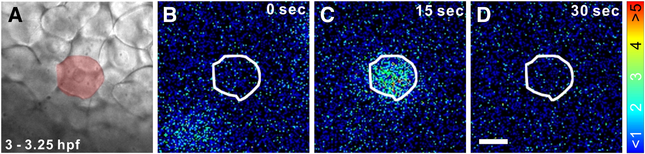
Fig. 5 Representative example of a Ca2+ transient that was generated during the dorsal-biased Ca2+ signaling window in a single SEC between 3.0–3.25 hpf. (A) A bright-field image showing a region of the superficial epithelium. The SEC that generated the Ca2+ transient is highlighted in red. Panels B to D show the representative pattern of aequorin-generated luminescence, in pseudocolor, illustrating the localized Ca2+ transient. The border of the SEC generating the transient is highlighted in white. Each panel represents 30 s of accumulated luminescence, and consecutive panels are stepped at 15 s intervals. The color scale indicates aequorin-generated luminescence in photons/pixel. Scale bar = 50 μm.
Reprinted from Developmental Biology, 327(1), Ma, L.H., Webb, S.E., Chan, C.M., Zhang, J., and Miller, A.L., Establishment of a transitory dorsal-biased window of localized Ca(2+) signaling in the superficial epithelium following the mid-blastula transition in zebrafish embryos, 143-157, Copyright (2009) with permission from Elsevier. Full text @ Dev. Biol.