
|
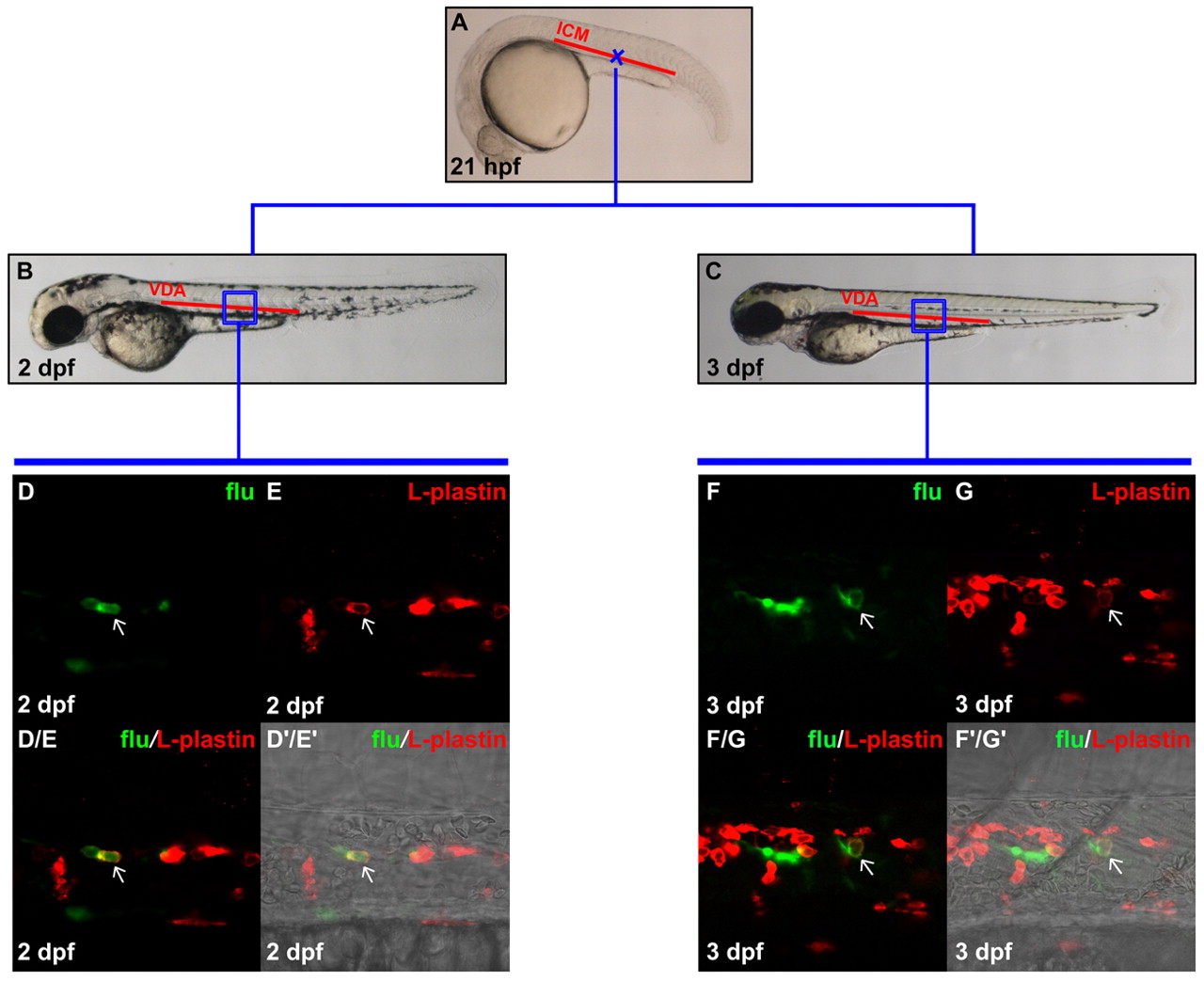
Fig. 4 Definitive myeloid cells are generated in the VDA. (A) Lateral view of 21 hpf embryo, indicating the uncaging position (blue cross) in the anterior part of the ICM. (B,C) Lateral view of 2 dpf (B) and 3 dpf (C) embryos. The boxed regions indicate the relative positions in the VDA where flu and L-plastin protein double-positive cells were detected after uncaging. (D,E) Confocal images of the boxed region in B showing the flu signal and L-plastin staining in the VDA at 2 dpf after uncaging at cross in A. (D/E) Merged image of D and E. (D′/E′) Superimposed view of D/E and DIC image. (F,G) Confocal images of the boxed region in C showing the flu signal and L-plastin staining in the VDA at 3 dpf after uncaging at cross in A. (F/G) Merged image of F and G. (F′/G′) Superimposed view of F/G and DIC image. Staining for flu and L-plastin is pseudopainted as green and red, respectively. Arrows indicate co-staining of flu and L-plastin.