
Image
|
Figure Caption
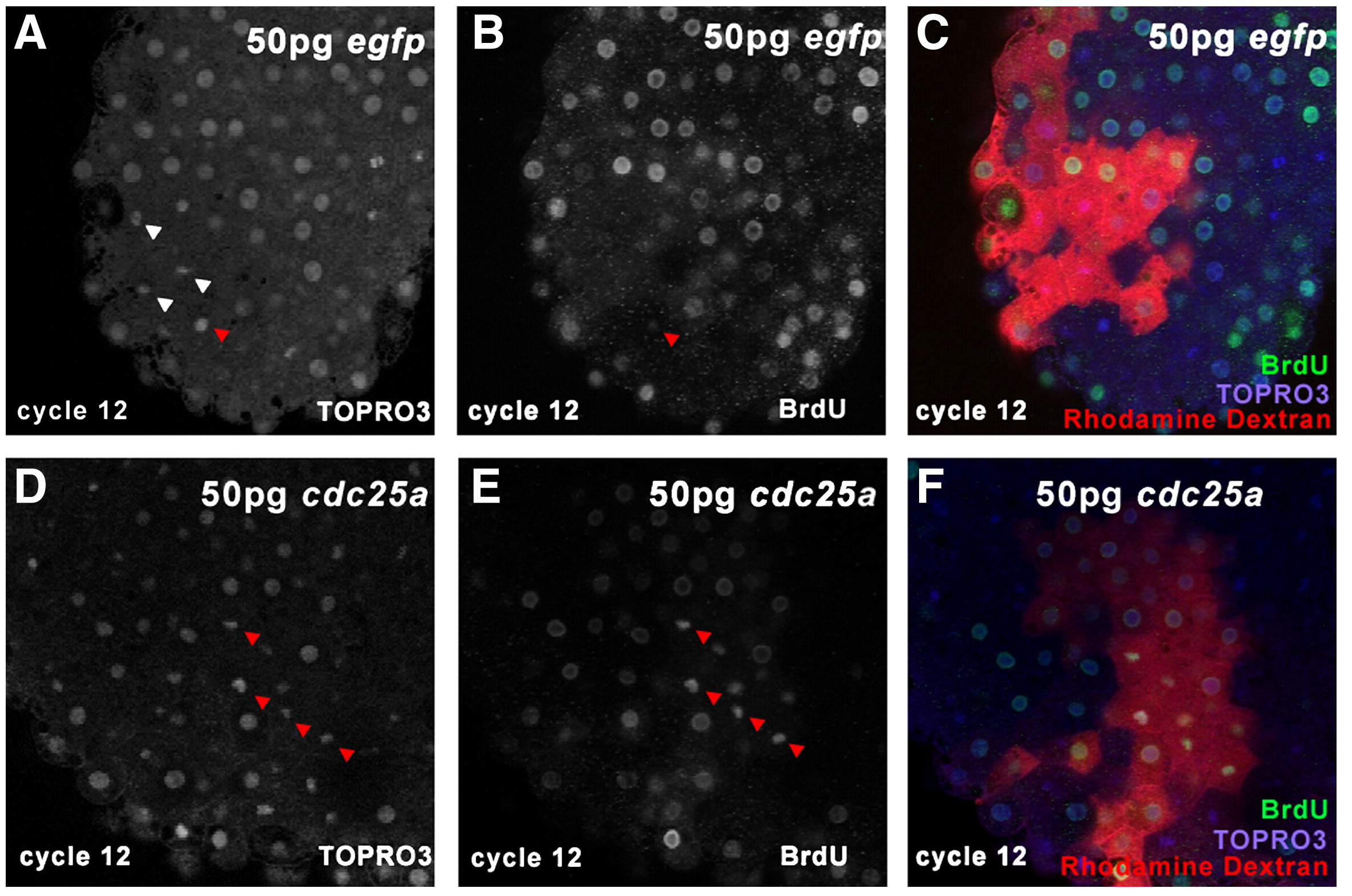
Fig. 6 BrdU mitotic pileup in clones injected with 50 pg egfp mRNA (A–C) or 50 pg 6MT-cdc25a mRNA (D–F). Embryos were injected with mRNA mixed with 70 K rhodamine dextran (C, F) then exposed to BrdU and nocodazole at 28 °C for 15 min. Embryos were then stained with anti-BrdU monoclonal antibody (white in B, E; green, C, F) and nuclei were counterstained with TO-PRO 3 (white in A, D; blue in C, F). Red arrowheads in panels A, B, D and E indicate BrdU-positive mitotic figures, and white arrowheads in A indicate BrdU-negative mitotic figures.
Acknowledgments
This image is the copyrighted work of the attributed author or publisher, and
ZFIN has permission only to display this image to its users.
Additional permissions should be obtained from the applicable author or publisher of the image.
Reprinted from Developmental Biology, 326(1), Dalle Nogare, D.E., Pauerstein, P.T., and Lane, M.E., G2 acquisition by transcription-independent mechanism at the zebrafish midblastula transition, 131-142, Copyright (2009) with permission from Elsevier. Full text @ Dev. Biol.