
|
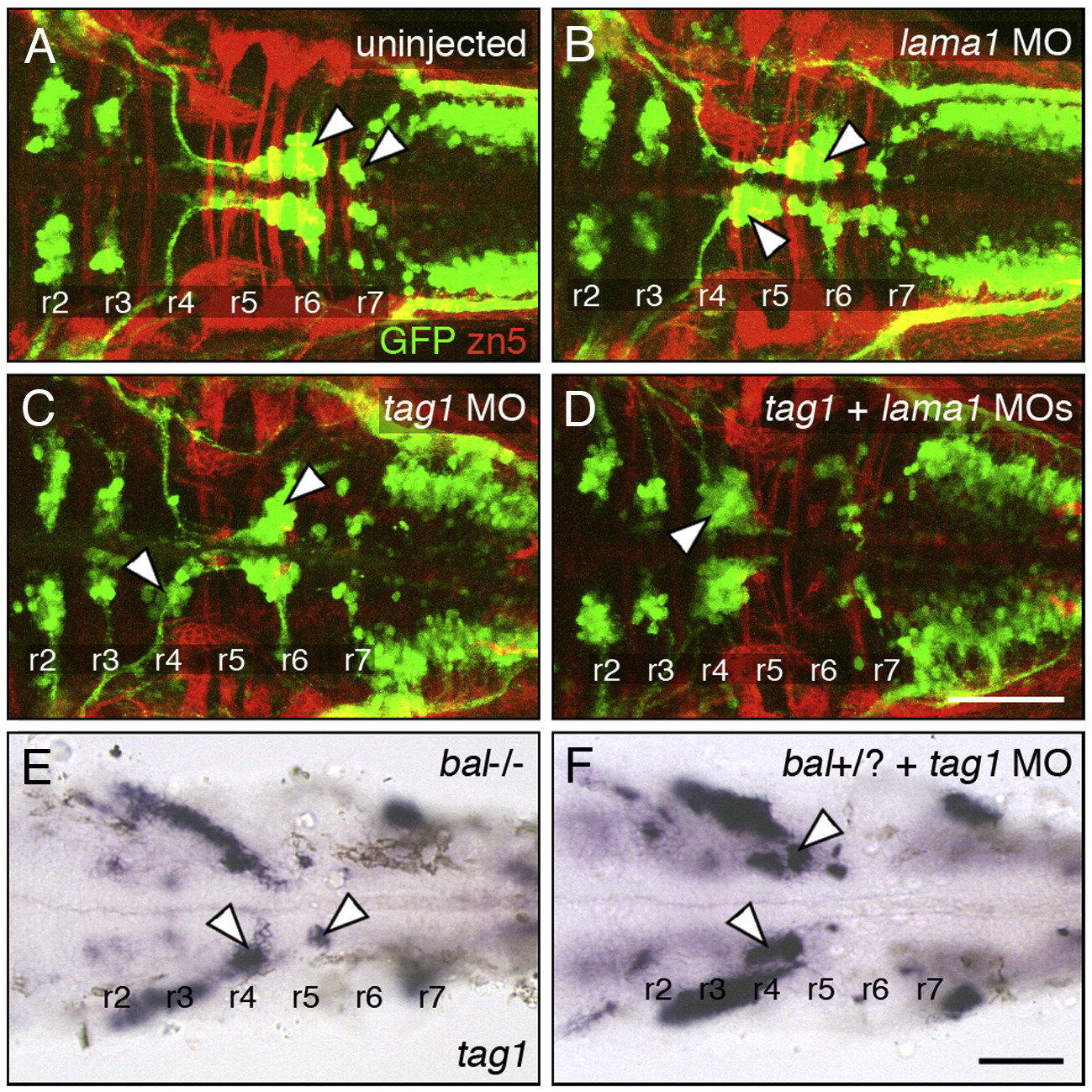
Fig. 7 Genetic interactions between tag1 and lama1. All panels show dorsal views of the hindbrain with anterior to the left. Tg(isl1:gfp) embryos (A–D) were fixed at 48 hpf, and processed for immunohistochemistry with zn5 antibody (red) to label dorsal commissural neurons and axons at rhombomere boundaries, and anti-GFP antibody (green) to label FBMNs (arrowheads). Non-transgenic embryos (E, F) were fixed at 30 hpf, and processed for tag1 in situ hybridization to label FBMNs (arrowheads). (A) FBMNs migrate normally in a control embryo. (B, C) Partial loss of FBMN migration in embryos injected with suboptimal dose of lama1 MO (B) or tag1 MO (C). (D) Complete loss of FBMN migration in an embryo injected with suboptimal doses of tag1 and lama1 MOs. (E) Greatly reduced FBMN migration in a bashfuluw1 homozygous (lama1 -/-) embryo. (H) Severe, nearly complete loss of FBMN migration in a putative bashful heterozygote injected with suboptimal dose of tag1 MO. Scale bar in D (75 μm for A–D); in F (75 μm for E, F).
Reprinted from Developmental Biology, 325(2), Sittaramane, V., Sawant, A., Wolman, M.A., Maves, L., Halloran, M.C., and Chandrasekhar, A., The cell adhesion molecule Tag1, transmembrane protein Stbm/Vangl2, and Lamininalpha1 exhibit genetic interactions during migration of facial branchiomotor neurons in zebrafish, 363-373, Copyright (2009) with permission from Elsevier. Full text @ Dev. Biol.